The Pressure Fallacy: What Pink Teeth in the Dead Really Reveal, and What They Do Not
A forensic investigation through 200 years of literature, prompted by three cases, two of which I examined myself, whose seemingly shared pattern I misinterpreted. About a rare postmortem finding that looks like a clue to the cause of death but is not. And about a mechanism that half the field keeps citing, even though no one ever proved it.
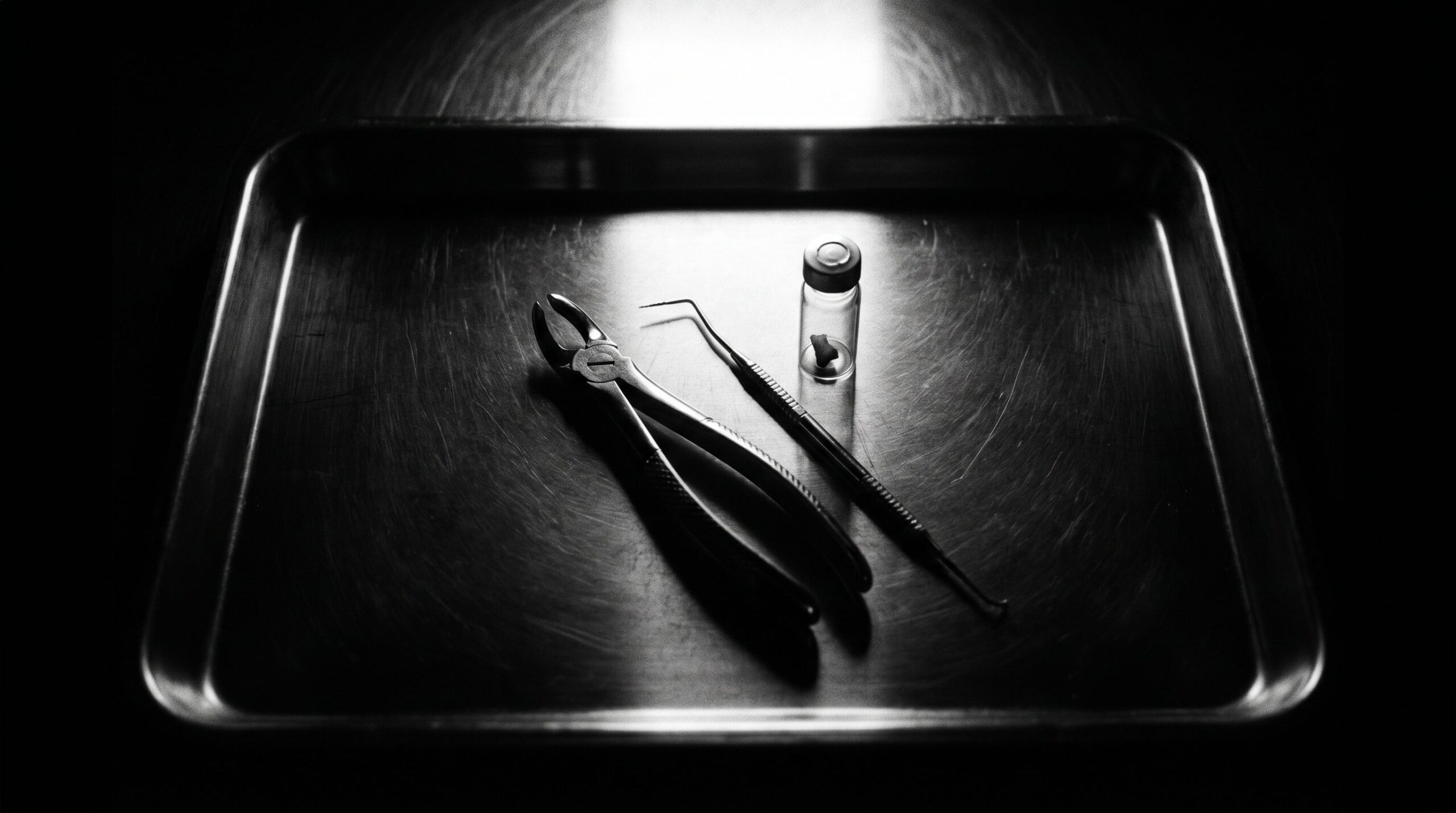
Hawaii, a Question at the Table, and My Wrong Answer
It was about five years ago. A multi-day advanced training in forensic anthropology with Professor Robert Mann in Hawaii, the topic was trauma. At some point a case appeared on the projection screen, a case from the past that we were discussing together, a person who had fallen from a building. The teeth of this individual showed a discoloration that no one in the room could quite place, a pink that did not rest on the surface but emerged from deep within the tooth, as though someone had saturated the dental hard tissue from the inside.
The question hung in the air: how does something like this come about?
I gave an answer. I said it was probably related to pressure. To a sudden, massive increase in pressure inside the skull. I did not say this at random, I said it from experience, and that is precisely what made the error so seductive. Nobody in the room knew better. We left the question open, the way one leaves questions open that one secretly believes have essentially already been answered. I threw the pressure hypothesis into the room and promised to look into the subject.
I am keeping that promise now. About five years late, but with the care it deserved from the beginning.
Three Cases, One Pattern, and Why That Was Exactly the Trap
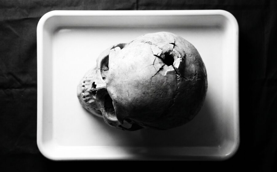
Three cases had lodged in my memory. I had worked two of them myself, a suicide by firearm and a fatal road accident with a shattered skull. The third was that fall from the building we had discussed on screen with Mann. Anyone who works cases looks for patterns, that is half the profession. And here the pattern was practically shouting at me.
Gunshot to the head, pressure. Frontal impact with skull fracture, pressure. Impact after a fall from height, pressure. Three deaths, three completely different causes of death, and yet a common denominator so clean it no longer looked like coincidence. A sudden, massive increase in pressure inside the skull. From this correlation I drew my conclusion. The pressure forces blood into the tooth, and that produces the color. Logical, right?
No, wrong. And the interesting thing about this story is that it is not about a novice getting something confused, but about how a perfect correlation deceives even a man who spent 25 years explaining to courts why correlation and causation are two different things. Three data points. One clear pattern. And the wrong mechanism behind it. If three cases are enough to convince me, then three cases are also enough to lead me astray.
What eventually brought me back to the subject was not a new case. It was research on a completely different topic, during which I stumbled across pink teeth again. I remembered them as rare, and that was correct. But I realized I had never actually known the cause, only ever confused my own plausible conjecture with the truth. So I sat down. Days of literature, from the first description in 1829 to review articles published this year. Plus a survey in my professional network, kept simple: what do you know about this, in which cases have you seen it? The answers were so heterogeneous that they already shook my pressure theory.
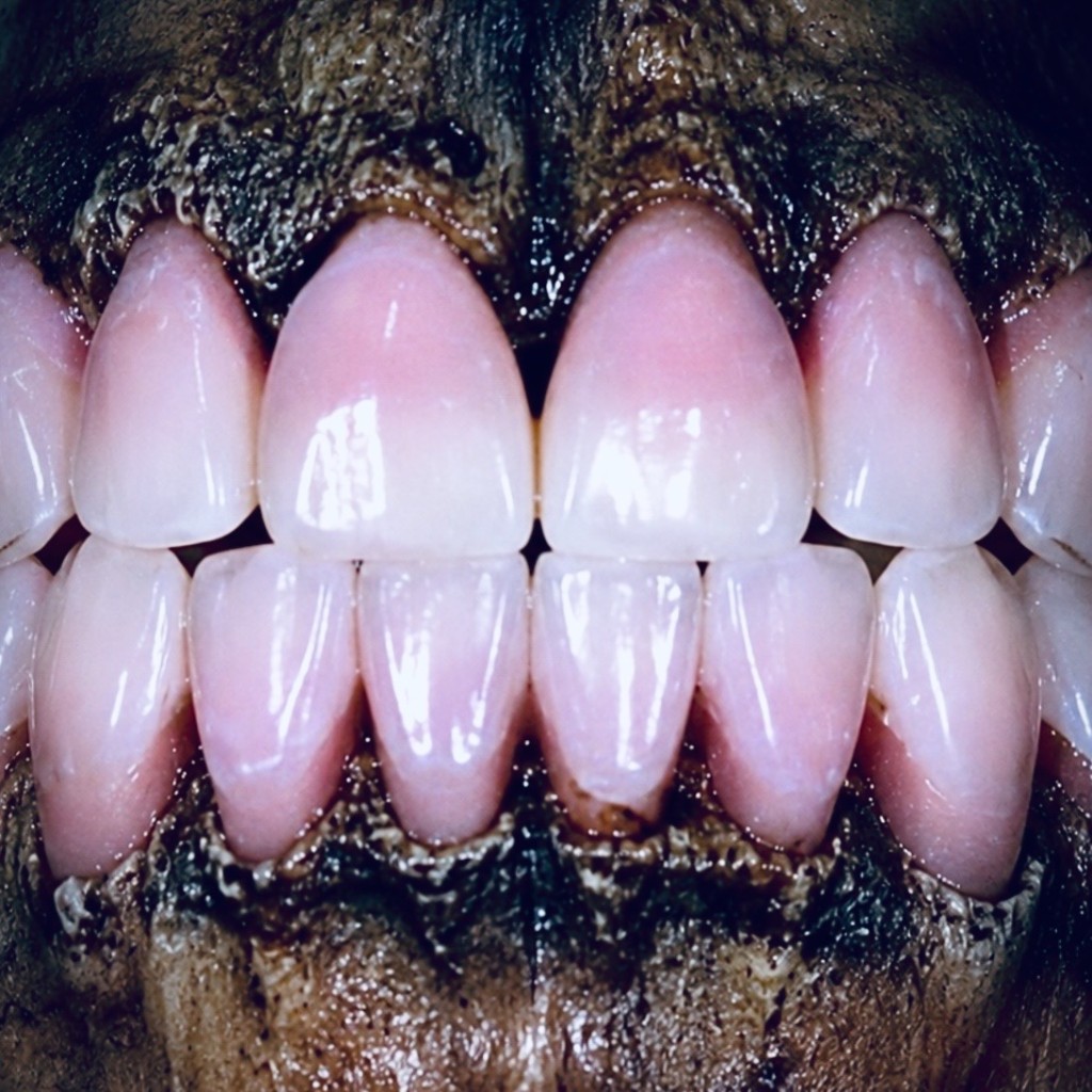
What Pink Teeth Are, and How Long We Have Known About Them
The phenomenon is old. It was first described by the English dental surgeon Thomas Bell in 1829. Bell examined the teeth of people who had died by hanging or drowning and found the dentine, the calcified tissue beneath the enamel, deeply reddish in color, while the enamel itself remained unaffected. Bell already had a hypothesis about the cause, and his hypothesis, remarkably, was: increased pressure inside the tooth. My error therefore has a distinguished ancestor. If you are going to be wrong, at least be wrong in the company of the person who first described the thing.
After that, nearly a century passed in silence. The subject vanished from the literature, until it returned in Japan in 1941. The researcher Kato published an extensive experimental series on cervical compression in 203 rabbits. Alongside it stood 4 human strangulation cases, not a comparable experiment but observations made at the autopsy table. Kato’s work was available only in Japanese for decades and was not systematically brought to an international audience until Sakurada did so in 2023. One point is important, and I will return to it: independently of Kato’s cervical compression experiments, pink teeth were later described in fire deaths, poisonings, gunshot injuries, natural deaths, and cases of unknown cause of death. The broader literature thus made it increasingly clear that the phenomenon is not confined to any single cause of death.
Forensic prominence came through a criminal case. London, 1953, the case of serial killer John Christie. Convicted and hanged solely for the murder of his wife Ethel, he is attributed with at least 8 killings between 1943 and 1953. His victims were usually incapacitated with coal gas and then strangled, a recurring pattern. And now comes the point at which I should already have paused, had I known the case more closely. Pink teeth were documented in only a single one of those bodies. Similar manner of death, yet the phenomenon appeared almost nowhere. The histochemical and spectroscopic analysis of that single finding was carried out at the time by Professors Miles and Fearnhead at the London Hospital. Already in 1953, that case provided the first indication that pink teeth are not a reliable sign of any particular cause of death. It was no more than a hint, because the fact that only one body showed pink teeth says nothing by itself about whether the other victims were systematically examined for the finding at all.
The Mechanism, and Why My Pressure Model Already Fails at the Level of Basic Anatomy
Now things get technical, and this is exactly where my old theory gets its first crack.
The dentine is traversed by tiny channels called dentinal tubules. They run from the pulp, the living dental tissue in the interior, outward toward the enamel. These tubules are widest near the pulp, where they measure about 2.5 micrometers, and they narrow toward the outer surface to less than 1 micrometer. Inside the pulp sits the blood, and in that blood float the red blood cells, the erythrocytes. A human erythrocyte measures roughly 7.5 micrometers. Hold those figures side by side. The channel is at its widest about 2.5 micrometers, the blood cell is three times that. An intact erythrocyte simply does not fit.
That means something fundamental. The size relationships clearly argue against intact red blood cells entering the dentinal tubules directly. Erythrocytes are remarkably deformable and pass through capillaries in the living circulation that are narrower than their resting diameter. This deformation is not an active effort by the cell, it results passively from membrane properties under a pressure gradient. The decisive difference lies elsewhere. Capillaries are compliant vessels lined with endothelium, dentinal tubules are rigid, mineralized channels. That a brief pressure impulse at the moment of impact forces intact erythrocytes directly into these rigid tubes has been neither experimentally demonstrated nor mechanistically plausible. The color requires something else, it requires hemolysis. My original model, that a pressure impulse at the moment of trauma forces whole blood cells directly into the dentinal tubules, was no longer tenable. Disproved was precisely that notion, the one I had never bothered to check.
What actually happens runs differently and above all more slowly. After death, the erythrocytes in the pulp begin to break down, they hemolyze. The cell membranes rupture, and hemoglobin, the red pigment, is released. Only now, as a dissolved molecule rather than a whole cell, can the pigment enter the narrow tubules at all and diffuse through the dentine. This is exactly why the whole process takes time. A person who has just died will not, as a rule, show pink teeth. The hemolysis must first occur, the pigment must then migrate, and that takes time. There are a few poorly documented reports of early discoloration appearing within the first one to two weeks, which I will return to at the end. The rule, however, is clearly a process that requires time.
Here the entire error of reasoning lies exposed. My model relied on a moment, on the fraction of a second of impact or gunshot. The real process is the exact opposite of a moment. It is a process over days to weeks, and what happens to the body after death determines far more about the coloration than the moment of dying. That does not mean the circumstances around the death play no role at all, blood distribution, congestion, and vascular condition can influence the available substrate. But the engine of the coloration starts running afterward.
The diffusion itself obeys the plain laws of physical chemistry, not the dramaturgy of a violent crime. Whether and how far the dissolved hemoglobin travels depends on the environment. Moisture keeps the pigment mobile and is the strongest known facilitator, even if it has not proven to be a strictly necessary condition in every documented case. Temperature governs the rate of decomposition, cold can preserve the red pigment longer but simultaneously slows hemolysis and diffusion, so the visible effect depends on timing and sequence. Oxygen and light work against the color, under air exposure it is weaker, under light it fades. This is not a random ensemble of factors, these are the classic variables of any diffusion in a porous material. Not one of these variables is set at the moment of death. All of them are adjusted afterward, by the earth, by water, by time.
Van Wyk demonstrated histochemically in 1989 that the staining pigment is undecomposed hemoglobin, and he noted that neither the cause of death nor the time of death had any observable influence on the histochemical detection reaction for hemoglobin in the teeth he examined. One could hardly bury my pressure theory more explicitly than that.
Which Pigment It Actually Is Remains a Matter of Debate
At this point, an honest admission that any serious treatment of this phenomenon must include. The question of exactly which hemoglobin derivative produces the pink color has not been definitively settled, and the answer depends on who you ask and what method they used.
Van Wyk arrived at undecomposed hemoglobin by histochemistry and UV microscopy and explicitly excluded hemosiderin, bile pigments, and porphyrins. Clark and Law, by contrast, isolated a protoporphyrin from pulverized pink dentine by thin-layer chromatography in 1984. Beeley and Harvey had already demonstrated hemoglobin and its haem derivatives through spectral studies in 1973 and even suggested an unstable complex with carbon monoxide. And the most recent controlled experimental model, a pilot study by Sumi and colleagues from 2023 using bovine teeth, found a spectroscopically rising carboxyhemoglobin signal after 7 days of decomposition, along with rising values for total haem and bilirubin over time. Crucially, and this is frequently swallowed in the secondary literature: this signal emerged postmortem in a model using hemolyzed bovine blood. It demonstrates a chemical change in the pigment after death, not a carbon monoxide poisoning during life. Confusing the two turns a postmortem chemical change in a laboratory model into a vital CO exposure.
Four studies, four slightly different answers. That is not a scandal, it is normal science. The findings broadly support hemoglobin and hemoglobin-related degradation products, but the precise chromophoric composition has not been established with finality. Which derivative dominates in any given case presumably depends on environment, temperature, and time, and part of the divergence between studies likely reflects differences in sample age, state of decomposition, and measurement method. Anyone expecting the Pantone number for death will be disappointed. Nature works with a blurred palette here, not a label.
The pilot study on bovine teeth produced a second finding that matters to me. Under controlled laboratory conditions the coloration was weaker under air exposure, faded under light, was more intense under the conditions tested at 4 degrees cold, and particularly reddish at high humidity. It peaked in the model after about 7 days and then faded again. That is a curve for bovine teeth in a laboratory setup, not a validated timetable for human remains, and it should be read as such. What remains: not one of these variables has anything to do with the cause of death. They all speak to what happens to the body after death, to storage conditions and environment.
How Science Actually Identified the Pigment
A claim about a pigment is worthless until someone has isolated and measured it. That is precisely what distinguishes the serious literature on this subject from guesswork, and it is worth pausing for a moment on the methods, because they show how hard people actually worked here.
Perhaps the most compelling evidence was provided by Kirkham and colleagues as early as 1977. They extracted the pink material directly from the dentine and demonstrated hemoglobin and serum proteins within it. Then they turned the argument around and reproduced the phenomenon experimentally by introducing blood into pulp chambers and allowing the teeth to mature under moist, decomposition-like conditions. What works in the laboratory with introduced blood requires no pressure impulse in nature, it requires only blood, moisture, and time. This reproduction is, for me, the real nail in the coffin of my pressure theory, because it demonstrates the phenomenon entirely without any mechanical impulse. Strictly speaking it proves that pressure is not necessary, not that vascular congestion can never contribute. But that pressure is required, as my old model claimed, is clearly ruled out.
The remaining methods read like a cross-section of analytical chemistry. Spectrophotometry and isoelectric focusing by Beeley and Harvey. Thin-layer chromatography by Clark and Law, who isolated their protoporphyrin that way. Histochemistry combined with UV microscopy by van Wyk, who pinned down the undecomposed hemoglobin. Electron beam microanalysis, which made the iron derived from hemoglobin visible directly within the dentine. And finally the modern colorimetric measurement in the Lab color space, with which Sumi and colleagues in 2023 quantified the color development over time, with very high inter-rater agreement.
What stands out in this methodological variety is an absence. For an independent, pressure-driven transport mechanism, no direct experimental evidence exists to this day. All measurement series capture pigment, iron, protein, color over time. The system under study is always the same: a pigment that, after death, slowly migrates from decomposed blood into the dentine. Active pressure never appears in any of these measurements as a variable in its own right, no one has ever pinned it down as a driving force.
The Mechanism Everyone Cites and Nobody Has Proved
Now I come to the part of my research that annoyed me most as a scientist and also fascinated me most. Because the pressure hypothesis that caught me out actually appears in the literature. Only it appears there in a very different place from what the citations claim.
One reads in numerous reviews that elevated intracranial pressure, venous congestion, and congestion of the head forces blood into the pulp and thereby promotes pink teeth. And usually this claim carries a reference to two classics, to Beeley and Harvey from 1973 and to Borrman and colleagues from 1994. That sounds solid. Two respected sources, one established explanation.
I checked. In the original texts of both those papers, this pressure explanation is not there. Beeley and Harvey describe biochemistry, spectra, haem derivatives. Borrman and colleagues describe no experimentally demonstrated pressure impulse forcing blood into the tooth. As preconditions for the subsequent pigment diffusion they discuss enhanced blood filling or congestion of the pulp, extravasation of erythrocytes, subsequent hemolysis, autolysis, and a moist environment, frequently water immersion. Congestion and extravasation do not exclude pressure-dependent vascular events, but nowhere is there any mention of a sudden pressure impulse that presses something into the tooth.
Where does the pressure formulation come from, then? In the sources I examined, the sharpened pressure formula can be traced back at least to a case report by Soriano and colleagues from 2009, a heavily decomposed body examined about a month after death, cause of death asphyxia. And Soriano grounded the pressure formula on Campobasso from 2006, not on Beeley and Harvey. From there the idea was passed along, often honestly labeled as hypothetical, and with each retelling it lost a little of its caution, until it looked like established knowledge traceable to Beeley, Harvey, and Borrman, even though none of them ever wrote it that way.
This is an instructive example of something I criticize often enough on this blog. A conjecture becomes a claim, the claim is cited, the citation becomes a fact, and eventually an entire discipline believes something that nobody ever proved. Otto Sapiens listens to an audiobook and knows it all. The specialist reads a secondary source and passes it on as primary knowledge. The difference is smaller than we would like. And I, who know exactly this, fell for the same mechanism, only my source was not a copied review but my own three cases. Self-deception has no sense of professional rank.
Moisture and Time, the Two Factors That Explain the Most
If not pressure, then what? The literature’s answer is unspectacular and for that reason convincing. It is the conditions after death, above all moisture and time.
Borrman and colleagues noted in 1994 that pink teeth, apart from a few poorly documented exceptions, develop at the earliest one to two weeks postmortem, and that a moist environment strongly promotes their formation, typically immersion in water. Van Wyk was able to reproduce the phenomenon in the laboratory and observed the pink discoloration macroscopically only after about six days, and even then only after hemolysis had begun. This fits a striking pattern in the literature. Many of the classic case reports involve water-recovered bodies, damp graves, and bodies that lay for extended periods in wet environments.
One case has particularly lodged in my memory. Campobasso and colleagues described in 2006 a series of bodies recovered from a single shipwreck, months after the sinking, long submerged. Many of these dead showed pronounced pink teeth. The discoloration was more pronounced in the younger victims. The reason lies in anatomy. Young teeth have wider, more permeable tubules and a larger pulp volume, the pigment has more room to travel. With age the tubules narrow and calcify, the pulp cavity shrinks, and the tooth becomes literally denser against the ingress of pigment. Secondary dentine, wear, restorations, and root canal fillings all work in the same direction. The phenomenon therefore does not favor a cause of death, it favors a tooth structure. The tendency is clear: people who die young are more likely to show pink teeth, regardless of what they died from. That is not a deterministic law of age, Minegishi found only a clustering below the age of 60. So much for the idea that the color reveals something about the end of a life. It reveals something rather about the architecture and age of the tooth.
Water immersion, moisture in the grave, long postmortem interval. These are the connecting lines, not intracranial pressure. And when I now set my three cases alongside this, I notice what I failed to see for five years. For not one of those cases had I documented what actually matters. How long had the deceased been lying until examination? Under what conditions of humidity? At what stage of decomposition? I had intracranial pressure in my head and never even noted the actually decisive variables.
What the Numbers Say, and Why They Bear Hard on My Theory
A theory can be refuted by mechanics, but the refutation only becomes convincing with numbers from real cases. And the results bear heavily on my original thesis.
The largest series to date comes from Minegishi and colleagues in 2022. They found pink teeth in 68 of 324 examined unidentified bodies, roughly 21 percent of this specific sample. This is not a general prevalence figure, since unidentified bodies are a highly selected group, often with long postmortem intervals and damp recovery environments. About 29 percent of all vital teeth were affected, most frequently the anterior ones, and markedly more often in individuals under 60. The decisive finding of this paper is a negative finding. There was no significant association between the presence of pink teeth and sex, place of discovery, or cause of death. A caveat is necessary here: the cause of death could actually be established in only part of the 68 positive cases anyway. The study thus shows no demonstrable association in a large but selected sample. It does not prove that such an association cannot exist in principle. For a reliable marker that can bear weight in court, however, exactly this distinction is decisive, and the finding does not support that use.
The systematic review by Franco and colleagues from 2019 pointed in the same direction. Across several studies, 71 bodies with pink teeth were found, the majority young adults, often recovered from moist environments, on average roughly three months after death. The authors’ conclusion is unambiguous. The phenomenon is nonspecific for cause of death.
Perhaps the most vivid counter-evidence was provided by a smaller paper by Brites and colleagues from 2020. The idea was elegantly simple. If pink teeth are a sign of asphyxia, as the old school claimed, they should appear with increased frequency in victims of asphyxia. The authors therefore looked deliberately at 21 victims of mechanical asphyxia, drowning victims, hanging victims, and one strangulation case. The result: a single case with pink teeth, a 26-year-old man who died by hanging. Roughly 5 percent. The sample is small, and a single positive finding in 21 cases does not disprove every conceivable vascular connection. But as what the old school claimed, namely a reliable marker for asphyxia, the finding in this series is clearly inadequate. The low hit rate speaks plainly against the forensic utility of the asphyxia thesis.
The Living Pink Tooth, a Completely Different Animal
Here I need to make a distinction that superficial accounts often pass over and that is forensically delicate. Because pink teeth also occur in the living. Only there they arise by an entirely different route and mean something completely different.
The living pink tooth is classically called the pink spot of Mummery, after the anatomist James Howard Mummery, who published a comprehensive study of these pink spots in 1920. The underlying phenomenon, internal resorption, was already described in the nineteenth century. What happens here is intravital, during life. Granulation tissue grows from the pulp outward, resorbing the dentine from within. Where the resorption has thinned the hard tissue sufficiently, the well-vascularized living tissue shows through the remaining shell as pink.
This is mechanistically the exact opposite of the postmortem finding. In the Mummery tooth the tooth is at least partially alive, the tissue is perfused, and the triggers are trauma, caries, an old restoration, or orthodontic treatment. A vitality test can return positive in the living patient, though it tests neural response rather than pulp vitality reliably, and in exhumed remains it is not applicable anyway. In the postmortem pink tooth, by contrast, the person is dead, the blood is decomposing, and the pigment migrates passively. Two entirely different events that happen by chance to share the same color impression.
Why does this matter forensically? Because the two can easily be confused in exhumed or heavily decomposed remains. Anyone who finds a pink tooth on a skeleton and reflexively concludes it is the postmortem phenomenon can be deceived by an old resorption that arose during life and has nothing whatsoever to do with the death. A clean differential diagnosis therefore belongs in any expert report that even mentions this finding. There is a third variant I mention for completeness, the tooth that turns grey or reddish after a blow during life because blood has entered the pulp. That too is intravital and does not belong in the same category.
The List of Confusions Grows the More Carefully You Look
Anyone who believes the differential diagnosis is finished once the living and dead pink tooth have been distinguished has stopped too soon. There are a number of other sources of reddish tooth discoloration, and an expert report that does not know them is vulnerable.
The most treacherous case is the one where the color does not come from blood at all. Dye and colleagues described archaeological pink teeth in 1995 and considered a fungal origin of the coloration probable. In long postmortem intervals, in skeletonized remains, in bone finds from the ground, this is a real possibility, and it has nothing to do with the hemoglobin-based phenomenon. Chromogenic bacteria that can form red pigments add to the list, though these tend to produce a surface biofilm rather than a pigment deep in the dentinal tubules, and the two must be distinguished. Heat exposure in fire deaths alters tooth color as well, depending on temperature and duration from yellowish through brown and grey to black. A specifically pink appearance from pure heat is not well documented, but the point belongs on the differential list, if only because fire and pigment can overlap.
On the intravital side the list may be even longer, though most of these findings differ morphologically from the classic cervical pink tooth. Tetracycline incorporation in childhood typically discolors multiple teeth in a banded yellow, grey, or brown pattern, not cervically pink. Congenital erythropoietic porphyria leads to erythrodontia, a reddish-brown discoloration of multiple teeth that fluoresces characteristically red under UV light. Certain dental materials, some older root filling materials, even physiologically resorbing deciduous teeth can give a pink impression. None of these findings says anything about the death, because they all arose during life. Anyone who sees a pink tooth at a scene and reflexively thinks of the postmortem finding can be badly wrong, and exactly that reflex is unacceptable in court.
The lesson is uncomfortable. The pink tooth is not an unequivocal sign, it is a question. The answer comes only from contextualizing it within postmortem interval, recovery environment, state of decomposition, and where appropriate targeted examination of the tooth itself, the dentine, extracted pigments, or, if still present, the pulp tissue. In advanced decomposition, assessable pulp tissue is often long gone, and the dentine is what counts. Anyone who confuses the question with a quick answer produces expert garbage with a scientific veneer.
What Remains of My Intuition, and It Is Not Nothing
At this point one might think my pressure theory has been thrown out entirely. It is not quite that simple, and the distinction here is to me the most important part of this whole text.
For a sudden pressure impulse as an immediate and independent cause of the color, there is no reliable evidence. Whether trauma as such is entirely irrelevant to the phenomenon is a question I deliberately leave open, and here I shift from finding to explicit hypothesis. An honest objection first, one that disarms me in advance: the living pulp is already well vascularized, blood is therefore present in abundance even without any trauma. For the subsequent hemoglobin diffusion, ruptured vessels are strictly speaking not required. That weakens my residual hypothesis before I even state it. What remains thinkable, no more: severe mechanical impact can rupture vessels and allow additional blood to extravasate into the pulp tissue, an intrapulpal hemorrhage. Theoretically, that could increase the available hemoglobin beyond what was already there and lower the threshold at which the phenomenon becomes visible. Theoretically.
That is a fine but decisive shift, and I need to label it precisely to avoid turning my old error into a new one. Pressure as the cause of coloration, that is wrong and settled. Trauma as an additional substrate provider, that is a hypothesis, nothing more. An independent, cleanly isolated association between skull trauma and the development of pink teeth has not been demonstrated, and it may well be that nothing of this remains in the end, because the pulp blood already present suffices. My three cases may share something, but the only certainty is what I never recorded, postmortem interval and moisture. Everything else I treat as what it is, a conjecture waiting to be tested.
The strongest concrete residual doubt about a model explained exclusively by advanced putrefaction comes from Kato and his reconstruction by Sakurada in 2023. Kato observed pink teeth in some cases early, before the actual onset of decomposition, following cervical compression. That is the clearest experimental indication that a vascular or mechanical contribution beyond putrefaction might exist. Honesty requires not suppressing this. But it is asphyxia by cervical compression, not blunt intracranial pressure, and it is a single, old dataset that urgently needs replication before anything is built on it. The remaining evidence is in any case heterogeneous and predominantly retrospective, and certainty exists nowhere in it.
To keep this from being mere opinion, I will state what would rehabilitate my old thesis. It would be rehabilitated once someone shows two things. First, pink teeth developing acutely, within hours of trauma, entirely without incipient putrefaction. Second, a statistically robust excess of pink teeth in skull trauma cases after postmortem interval and moisture have been properly controlled for. Until both of those are shown, the current evidence predominantly supports the multifactorial postmortem model of hemolysis, pigment diffusion, tooth structure, time, and environmental conditions, and my pressure idea remains what it is, a disproved hypothesis with an honorable ancestry. That is how one must treat one’s own assumptions if one wants to do science rather than profess faith.
A Warning to Everyone Who Takes This Finding into Court
Before I close, a word to colleagues, and I mean forensic pathologists explicitly, because there is a genuine need for clarification here.
Anyone who mentions pink teeth in an expert report should know precisely what they are entitled to say and what they are not. Defensible is the statement that hemoglobin-containing material from the pulp has migrated into the dentine, and that this process is typically promoted by time and a relatively moist environment. No more than that, and even here caution is warranted. That the pulp once contained blood says nothing by itself, since the living pulp is always vascularized. Moisture is a facilitating factor, not a condition that the finding would prove. And a firm minimum postmortem interval cannot be derived from pink teeth, the typical range starts at one to two weeks or later, but there are documented earlier individual cases that break every rigid lower bound. Not justifiable is the inference from pink teeth to a specific cause of death. Not justifiable is the claim that a pressure impulse inside the skull produced them. Not justifiable is the confusion with intravital resorption. And not justifiable is the use of the finding as a reliable marker for time of death, because the timeframe is far too broad and far too dependent on the environment for that purpose.
The medico-legal hazard is real. An expert who infers asphyxia or a specific sequence of events from pink teeth is building their conclusion on sand, and a good defense counsel will take it apart. The finding has its place, but only as one tile in a mosaic alongside autopsy, recovery circumstances, state of decomposition, and dental history. On its own it carries nothing. Letting it carry something on its own risks a miscarriage of justice and the expert’s own credibility.
The Promise Kept in Hawaii
Which brings me back to the person who fell from the building, and the question at the projection screen with Professor Robert Mann that remained open for five years.
I had thrown the pressure thesis into the room and promised to look into the subject. Today I know that the pink coloration of that person’s teeth proved nothing in itself about the fall, and nothing about a pressure impulse at the moment of impact. It showed that hemoglobin-containing material had entered the dentine, nothing more. What role postmortem interval, moisture, temperature, and decomposition played in exactly that case I can no longer reconstruct in hindsight, because I had not recorded the decisive variables at the time. That is the real point of the whole story. The most probable explanation lies in hemolysis, time, and postmortem environmental conditions, not in a momentary pressure impulse. But what I believed I knew for five years cannot be established with certainty for that one case, for the precise reason that I never collected the evidence that would have made it certifiable.
It is a small humiliation and a large satisfaction at once, to dismantle one’s own conviction cleanly after five years. The humiliation, because I was wrong, and wrong with the full weight of my own experience behind me. The satisfaction, because that is precisely the job. Not to defend an assumption but to test it, and to let it fall when the data do not support it. Three correlating cases seduced me. Two hundred years of literature corrected me. As it should be.
Bob, the literature review I promised you at the projection screen five years ago took five years. Here it is, late, and without pressure.
On exactly this subject I will write a detailed scientific paper that combines a systematic literature review with my own standardized documented case series, going substantially beyond the preliminary version presented here. For that I depend on material that exists in no database, but in the archives and on the hard drives of colleagues. Anyone who has documented pink teeth themselves, in whatever case, I am genuinely grateful for the submission of case and image material. Particularly valuable are cases with information on age, tooth position, postmortem interval, recovery environment, refrigeration, and state of decomposition, along with unedited original images with scale bar and color chart. The more such cases come together, the more robust the final statement will be. A single finding seduces, as mine did. A collection of well-documented cases clarifies.
References
- Bell, T. (1829). The Anatomy, Physiology, and Diseases of the Teeth. Highley, London.
- Aquila, I., Gualtieri, S., Princi, A., & Sacco, M. A. (2025). A systematic review about postmortem pink teeth: Forensic classification, diagnostic value, and analysis methods. Diagnostics, 15(16), 2092.
- Beeley, J. A., & Harvey, W. (1973). Pink teeth appearing as a post-mortem phenomenon. Journal of the Forensic Science Society, 13(4), 297–305.
- Borrman, H., Du Chesne, A., & Brinkmann, B. (1994). Medico-legal aspects of postmortem pink teeth. International Journal of Legal Medicine, 106(5), 225–231.
- Braga, S., Caldas, I. M., & Dinis-Oliveira, R. J. (2025). Forensic significance of postmortem pink teeth: A narrative review. Archives of Oral Biology, 169, 106092.
- Brites, A. N., Rezende Machado, A. L., Franco, A., & Alves da Silva, R. H. (2020). Revisiting autopsies of death by mechanical asphyxia in the search for post-mortem pink teeth. Journal of Forensic Odonto-Stomatology, 38(1), 34–38.
- Campobasso, C. P., Di Vella, G., De Donno, A., Santoro, V., Favia, G., & Introna, F. (2006). Pink teeth in a series of bodies recovered from a single shipwreck. American Journal of Forensic Medicine and Pathology, 27(4), 313–316.
- Clark, D. H., & Law, M. (1984). Post-mortem pink teeth. Medicine, Science and the Law, 24(2), 130–134.
- Dye, T. J., Lucy, D., & Pollard, A. M. (1995). The occurrence and implications of post-mortem ‘pink teeth’ in forensic and archaeological cases. International Journal of Osteoarchaeology, 5(4), 339–348.
- Franco, A., de Oliveira, M. N., Gomes-Lima, L. K., et al. (2019). Case-specific characteristics of pink teeth in dental autopsies: A systematic review. Journal of Forensic and Legal Medicine, 68, 101869.
- Franco, A., Mendes, S. D. S. C., Picoli, F. F., Rodrigues, L. G., & Silva, R. F. (2018). Forensic thanatology and the pink tooth phenomenon: From the lack of relation with the cause of death to a potential evidence of cadaveric decomposition in dental autopsies: Case series. Forensic Science International, 291, e8–e12.
- Kirkham, W. R., Andrews, E. E., Snow, C. C., Grape, P. M., & Snyder, L. (1977). Post-mortem pink teeth. Journal of Forensic Sciences, 22(1), 119–131.
- Lopes Cardoso, I., Sá, M., Moreira, M. T., & Guimarães, M. I. (2026). Postmortem pink teeth in forensic medicine: A scoping review of forensic significance and interpretive limits. Forensic Sciences, 6(2), 32.
- Miles, A. E. W., Fearnhead, R. W., Harrison, J. A., & Nickolls, L. S. (1953). Pink teeth. In F. E. Camps (Ed.), Medical and Scientific Investigations in the Christie Case. Medical Publications, London.
- Minegishi, S., Utsuno, H., Ohta, J., et al. (2022). Sixty-eight cases of postmortem pink teeth observed in dental autopsies of unidentified cadavers. Journal of Forensic Sciences, 67, 1280–1287.
- Mummery, J. H. (1920). The pathology of ‘pink-spots’ on teeth. British Dental Journal, 41, 301–311.
- Sakurada, K. (2023). Effects on oral tissues of asphyxiation caused by cervical compression: The pink teeth phenomenon in Kato’s studies (1941). Legal Medicine, 64, 102284.
- Soriano, E. P., Carvalho, M. V., Santos, F. B., Mendoza, C. C., Araújo, M. D., & Campello, R. I. (2009). The post-mortem pink teeth phenomenon: A case report. Medicina Oral, Patología Oral y Cirugía Bucal, 14(7), E337–E339.
- Sumi, N., Minegishi, S., Ohta, J., Utsuno, H., & Sakurada, K. (2023). Study on the mechanism of the pink tooth phenomenon using bovine teeth: A pilot study. Diagnostics, 13(16), 2699.
- van Wyk, C. W. (1988). Postmortem pink teeth: In vitro production. Journal of Oral Pathology, 17(9–10), 568–572.
- van Wyk, C. W. (1989). Postmortem pink teeth: Histochemical identification of the causative pigment. American Journal of Forensic Medicine and Pathology, 10(2), 134–139.
The Hole Lies, the Rim Tells the Truth: What a Photograph of a Shattered Skull Really Reveals
Forensic analysis of gunshot wounds to the human skull reveals that the hole is the least informative part…

The Skeleton Does Not Lie
What a skull on the piano gives away about a stranger's life, why bone is more honest than…

Otto Sapiens, the Last Model Before Extinction
A diagnosis of the Stone Age brain under the screen, the voluntary surrender of thought, and the single…