Mysterious Pupil Dilation in Comparison Photos: Stress or Drugs?
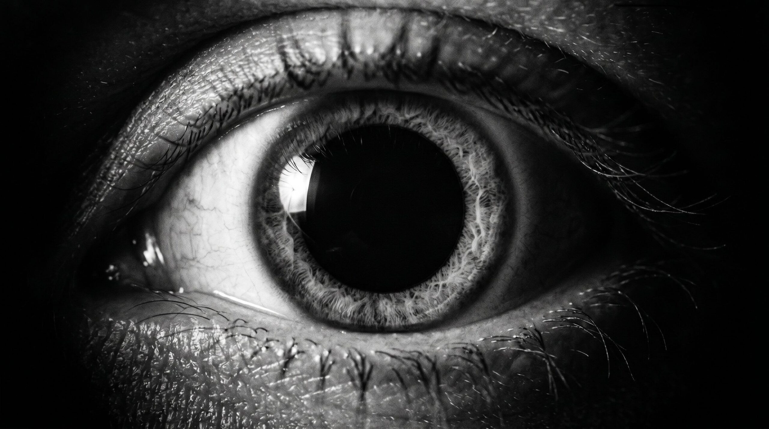
At the start of my work as an expert witness, the same thing kept catching my eye in the comparison photographs of defendants. Enormous pupils. Black discs that all but swallowed the iris, sometimes even under the light of a flash, that is, under an amount of light at which a healthy pupil ought to shrink to a small point. These images came almost exclusively from investigators or from the court. They were not party snapshots, not furtive pictures from some long night out, but sober official photographs taken in interview rooms and in the corridors of justice buildings. And still, eyes stared back at me that looked as though their owner had just taken something.
It would not let me go. A photograph in a criminal case is never merely a photograph. It enters the file, it is looked at, it creates an impression, and the impression of a wide open pupil whispers a single word to the viewer who does not know better. Drugs. That whisper is dangerous, because it translates a medical observation into a moral verdict without a shred of evidence in between. So I started digging, and what I found is more complicated, and at the same time clearer, than the quick answer suggests. One detail still nags at me. A photograph cannot be cross examined. A witness who claims the defendant had glassy eyes has to explain what he based that on, and defense counsel can press him. The photograph, by contrast, claims nothing and seems to prove everything. It simply lies there and works, and an impression that needs no words is harder to argue against than a stated assertion. That is exactly why I trained myself to be especially suspicious of these images.
The quick answer would in fact be stress. A large pupil is very often an ordinary stress response. But the quick answer is only the beginning, not the end. Before we talk about drugs, about stress, about rare eye conditions and about the patrol officer’s flashlight test, we have to understand briefly what a pupil even is. Not as a poetic window to the soul, but as what it mechanically really is. A valve.
The pupil is not a mood barometer but an autonomic valve
The pupil is nothing more than the hole in the middle of the iris through which light enters the eye. Its size is set by two muscles working against each other, and these two muscles obey two different masters. The sphincter, the musculus sphincter pupillae, narrows the pupil and is under parasympathetic control, the part of the autonomic nervous system responsible for rest, digestion, and recovery. The dilator, the musculus dilatator pupillae, pulls the pupil open and obeys the sympathetic system, the one that puts the body on alert. Steinhauer and colleagues teased this dual control apart in a controlled study and showed that pupil dilation comes about both through firing up the sympathetic system and through releasing the parasympathetic brake (Steinhauer et al., 2004).
The dilator is driven by norepinephrine, which docks onto so called alpha-1 receptors. That is the same messenger that makes the heart beat faster, raises blood pressure, and widens the bronchi. A wide pupil is therefore not a freestanding phenomenon, it is part of a whole program. The body switches to fight or flight, and the pupil is thrown open because more light should reach the eye, because the visual field should be sharpened, the threat fixed, the environment scanned with the highest attention. Evolutionarily this makes sense. Whoever faces the saber toothed tiger needs no pupil tuned to reading distance, but one that registers every movement at the edge of the field.
The pathways behind this are two separate lines. The parasympathetic command to constrict springs from a nucleus in the brainstem, the Edinger-Westphal nucleus, and runs with the third cranial nerve to the sphincter. The sympathetic command to dilate takes an absurdly long detour, descending from the hypothalamus down into the upper spinal cord and from there back up through a chain of relay stations in the neck to the eye. For our purposes only the consequence matters. Two independent systems pull on the same pupil, and its size at any given moment is always the result of a tug of war, never of a single command. This also explains why the photographs taken despite a flash troubled me so much. A flash is a strong light stimulus that ought to make the pupil contract. If it stays wide anyway, then in that moment the sympathetic dilation or the parasympathetic inhibition pulled hard enough that even a direct light stimulus could not overcome it. That tells me the arousal was considerable, and it still does not tell me where it came from. There is also a sober measurement problem that everyone in my field knows. The absolute size of a pupil cannot be reliably determined from an ordinary photograph at all. Without a scale in the image, without known lighting, without a defined distance, the viewer estimates, and he estimates against the background of what he already expects. A pupil one takes to be strikingly wide may simply have been photographed in a darkened room, where every healthy pupil is wide. The impression of a drug pupil then arises not in the eye of the photographed person, but in the head of the viewer. This is exactly where the line between observation and projection blurs, and exactly where an expert has to be most watchful.
This leads to something decisive for forensic assessment. The pupil is an indicator of the state of arousal, not of the cause of that arousal. It tells you the sympathetic system is firing. It does not tell you why it is firing. And this is precisely where the reflex of attaching the label drug to a wide pupil in an official photo goes wrong. The sympathetic system does not distinguish between a chemically produced fire and a fire produced by mortal fear. For the dilator muscle, norepinephrine is norepinephrine, whether a substance released it or the simple fact that the person is sitting in an interview room and does not know whether he will leave the building a free man.
The obvious suspect is the wrong one
What puts a person under more stress than an ongoing criminal investigation? I can think of little. An arrest, a search, an interrogation, the moment the photograph is taken, all of these are exceptional states in which the body runs exactly the program that throws the pupil open. The defendant need not have consumed the slightest thing. It is entirely enough that he is afraid, and in this situation just about everyone is afraid, the guilty and the innocent alike. And it need not even be fear. Steinhauer and colleagues showed that the pupil responds to arousal as such, regardless of whether the triggering feeling is pleasant or unpleasant (Steinhauer et al., 2004). Anger, tension, agitation, even the sheer sensory overload of a strange and intimidating environment drive the same sympathetic response. The pupil is an arousal meter, not a fear meter and certainly not a guilt meter. A person who feels wrongly accused and is boiling with indignation has wide pupils for the same reason as one who is trembling with fear. The image cannot tell the two apart, and the viewer who has only the image cannot either. This very ambiguity forbids any quick reading, and it vanishes the moment someone looks at the photo with a ready made expectation.
Bitsios and colleagues worked out a detail one has to know when assessing official photos. They had subjects anticipate an aversive event, specifically an electric shock, and measured the pupil during it. The result was clear. The mere anticipation of something unpleasant, before anything happened at all, increased the baseline diameter of the pupil and at the same time dampened its response to light (Bitsios et al., 2004). Anticipation is enough. The body need not actually be threatened, it need only see the threat coming. Translate that into the situation at the police station, and you have the explanation for a considerable share of the wide pupils that have crossed my desk on comparison photos over the years. The person sits there, expects nothing good, and his autonomic nervous system sets the pupil exactly as a stimulant would. One has to keep two speeds of the stress response apart here. The fast one runs purely neurally, the sympathetic system fires within fractions of a second, and the pupil reacts almost at once. The slower one runs hormonally through the adrenal gland and cortisol and takes minutes. On a comparison photo we see the fast, neural one, and that is exactly the one that widens the pupil. The person therefore need not have built up fear for hours, the moment of the photograph is entirely enough.
Here lies the real punchline, and it carries an irony that can spoil your appetite. The photographic situation itself produces the supposed drug sign. The photo meant to ground the suspicion produces the very feature on which the suspicion rests. You photograph a frightened person under stress and then read off the photo that the person is on something. In that moment the pupil’s sphincter has no chance against the sympathetic system, and the sympathetic system is running because the situation is what it is. Whoever does not know this mistakes the effect of the interrogation for the cause of the interrogation.
I do not want to pretend that this explains away every wide pupil. That would be the same short circuit in the other direction. Stress is the most common explanation, but not the only one, and a serious examiner does not make it easier than it is. Drugs can indeed be the cause. Just not all of them, and not as unambiguously as the gut would like.
When it really is drugs
Let us talk about the chemistry, and let us talk precisely, because this is where the greatest nonsense gets spread. There is no such thing as the drug that makes pupils large. There are classes of substances that pull in opposite directions, and that direction is the forensically decisive thing.
Stimulants widen the pupil. Amphetamine, methamphetamine, and cocaine act sympathomimetically, that is, they mimic the very fight or flight program that stress also switches on, by raising the availability of norepinephrine and dopamine in the nervous system. Holze and colleagues showed in a controlled double blind study with 28 healthy subjects that d-amphetamine, alongside LSD and MDMA, triggers a clear sympathomimetic response that includes a measurable increase in pupil size (Holze et al., 2020). The study worked with clearly defined doses, 40 milligrams of d-amphetamine, and compared them directly against 0.1 milligrams of LSD and 125 milligrams of MDMA, with all three substances widening the pupil to a comparable degree (Holze et al., 2020). Cocaine adds a local anesthetic component, but changes nothing about the basic sympathomimetic effect on the pupil. The pupil responds to cocaine or methamphetamine no differently in principle, because the mechanism is the same.
MDMA, the ecstasy of club nights, belongs in its own drawer but strikes the same note. Hysek and Liechti used infrared pupillometry to measure exactly what the substance does to the eye. MDMA produced a mydriasis, that is, a wide pupil, lengthened the reaction time to light, and weakened the light response, and this effect lasted as long as the substance circulated in the blood and only normalized after about 6 hours (Hysek & Liechti, 2012). Unlike with pure stimulants, this is mediated by an interplay of norepinephrine and serotonin. This measurement was no incidental observation, but came from five placebo controlled studies with 16 healthy subjects each, in which the pupil was recorded by infrared pupillometry (Hysek & Liechti, 2012). Such controlled conditions are the reason one can trust the finding, unlike the everyday observation in the interview room, where dozens of confounders run together.
The hallucinogens, finally, LSD, psilocybin from the mushroom and mescaline from the cactus, widen the pupil by a third route. They act on the serotonin system, specifically at the 5-HT2A receptor, which Halberstadt describes in his review as the primary site of action of this class of substances (Halberstadt, 2015). That this serotonergic attack reaches all the way to the pupil is shown again by the Holze study, in which LSD widened the pupil just as MDMA and amphetamine did (Holze et al., 2020). Three different chemical routes, one shared visible result.
And now the part that regularly drops out of the reflexive equation wide pupil equals drug. There is a large, socially highly relevant class of substances that does the exact opposite. Opioids, that is, heroin, morphine, fentanyl, and the whole family, constrict the pupil down to pinhead size. Whoever infers drug use from a wide pupil thereby practically rules out opioids and points at most to stimulants, MDMA, or hallucinogens. The pupil is therefore not the lie detector for consumption as such, but at best a crude hint at a particular direction. It can open one door and close another, but it never names the substance. In emergency medicine this is captured under the term toxidromes, the typical symptom combinations of individual substance groups. The sympathomimetic toxidrome of the stimulants goes with wide pupils, the opioid toxidrome with pinhead sized ones. Whoever knows only the pupil and nothing else can at best roughly narrow down a toxidrome, but never name a specific substance, and certainly never prove consumption. There is a second dimension the photo conceals, time. A stress related widening is fleeting, fluctuating second by second with the inner state and gone as soon as the person calms down. A substance related widening, by contrast, persists as long as the substance circulates in the blood, and with MDMA the pupil width even follows the substance’s concentration curve and only recedes after hours (Hysek & Liechti, 2012). A single photo, however, is a snapshot without a time axis. It cannot distinguish between the fleeting stress moment and the lasting substance effect, because it freezes both onto the same single instant. Whoever derives a statement about hours from that one frozen second attributes to the image information it technically does not contain.
The medical doppelgangers
There is a third group of causes that gets readily overlooked in the heat of suspicion, because it is neither spectacular nor morally charged. A wide or poorly reacting pupil can simply be a physical finding that has nothing whatsoever to do with stress or drugs. Whoever does not have this on the radar mistakes a congenital or acquired anatomy for proof of guilt.
Let us begin with the most extreme case, aniridia. Here the iris is wholly or almost wholly absent from birth, usually through a change in the PAX6 gene, so that the eye looks as though it consists of nothing but one enormous pupil. Landsend and colleagues describe the whole range of this rare disorder in their comprehensive review, with a prevalence between 1 in 64,000 and 1 in 96,000 people (Landsend et al., 2021). Rare, to be sure, but whoever meets such a person and reflexively bets on drugs makes a thorough fool of himself, because here the seemingly gigantic pupil is simply the absence of the iris.
Then Adie’s syndrome, also called the tonic pupil. Here one pupil is permanently wider than the other and reacts only sluggishly and with delay to light, because the parasympathetic supply to the sphincter is damaged. Kawasaki describes this and related disorders of pupillary function at length in the standard neuro-ophthalmology literature (Kawasaki, 2005). Such a pupil is indistinguishable on a photo from a drug related mydriasis if one does not know the history, and the history simply is not printed on the photograph. Then there is traumatic mydriasis. A blunt blow to the eye, a fall, an accident, and the fine ring muscle of the iris tears. The pupil then stays permanently wide and no longer reacts properly, because the muscle that should narrow it is mechanically damaged. This too is described in the standard neuro-ophthalmology literature as a tear of the iris sphincter with subsequent pupil dilation (Kawasaki, 2005). A person whose eye was injured ten years ago in a brawl or in traffic carries this wide pupil as a scar, entirely sober, entirely unremarkable otherwise. Add to that the utterly banal cases, such as eye drops with an anticholinergic effect, of the sort the ophthalmologist uses to examine the back of the eye, which set the pupil wide for hours. Whoever visited the eye doctor shortly before the photo looks like a user on the image and is only a patient.
There is a distinguishing feature here that is worth its weight in gold in practice and is nonetheless constantly overlooked. Stress and drugs act on both eyes equally, they widen both pupils symmetrically, because they act through the bloodstream or through the central nervous system on both sides at once. A one sided wide pupil, a so called anisocoria, therefore argues precisely against drugs and against stress and for a local or neurological cause, an Adie’s syndrome, an old injury, a pressure injury to the third cranial nerve (Kawasaki, 2005). Whoever sees two differently sized pupils on a photo and thinks of drugs has overlooked the obvious, because no stimulant on earth preferentially widens the left eye. Symmetry is the first question one has to ask, and it is almost never asked. One special case deserves particular attention, because it can be literally life threatening. A one sided wide, light fixed pupil can be a sign of pressure on the third cranial nerve, for example from a bleed or a swelling in the skull (Kawasaki, 2005). That is not a drug problem, that is a neurosurgical emergency. Whoever reads such a person’s pupil as a sign of consumption instead of getting him to a hospital commits an error that goes far beyond a wrong note in a file. The first duty toward a striking pupil is therefore not the criminological one, but the plain medical question of whether someone here needs help.
The flashlight test and its limits
In practice the patrol officer makes do with a simple tool that, for all its modesty, is remarkably informative. He shines a lamp into the suspect’s eye and watches what the pupil does. That is high tech diagnostics with hardware store means, and it works because it does not check the size of the pupil, but its reaction.
The underlying mechanism is the pupillary light reflex, and it runs through the parasympathetic system. When bright light hits the retina, the sphincter is instructed to narrow the pupil promptly. In a sober, healthy person this happens quickly and forcefully. Under the influence of certain substances, by contrast, this reaction is slowed, weakened, or absent altogether. Hysek and Liechti measured exactly that for MDMA. The substance lengthened the latency to the reaction and reduced the constriction to light, a finding that makes the central parasympathetic inhibition visible (Hysek & Liechti, 2012). Bitsios too showed that the amplitude of the light reflex changes with the inner state, that not only the resting size but precisely the reaction is a sensitive indicator (Bitsios et al., 2004). A sluggish or absent light reaction is therefore often more telling than a merely wide pupil. The reflex itself is an elegant arc. The light hits the retina, the information runs over the optic nerve to the brainstem, is switched there onto the parasympathetic nucleus, and returns over the third cranial nerve to the sphincter. If only one eye is illuminated, both normally constrict, the illuminated one and the other, because the wiring in the brainstem couples both sides. This coupled reaction alone gives the trained observer more information than the bare pupil size, because it tests a whole conduction pathway at once.
But, and this but is large, the test is a quick screen and not a proof. A sluggish light reaction is also found in Adie’s syndrome, after eye injuries, in great exhaustion, and under medications that have nothing to do with illegal substances. And conversely, an intact light reaction does not safely rule out consumption. The flashlight test narrows the suspicion, it does not confirm it. Whoever turns it into a diagnosis overstretches a tool that is good for a first assessment and not fit for a court proof finding. For the latter you need a blood or urine analysis, that is, the detection of the substance itself, and not the interpretation of a muscle that can stand wide for a dozen reasons.
Why this matters in court
One might take all this for a medical nicety, for knowledge that is nice to have and nothing more. That would be a mistake. In a criminal case the impression weighs in, and a comparison photo with wide open pupils creates an impression long before anyone has read a toxicological report. The viewer, be it a lay judge, an investigator, or a journalist, sees the black discs and thinks what he is conditioned to think. There is someone who has taken something. Out of this silent assumption comes a context, and the context colors the assessment of every further piece of evidence.
This is exactly where it is my job as a forensic examiner to pull the brake. Not because I want to shield users, but because a stress response is not a confession and must never pass for one. Over the years I have seen enough of these photos to know that the wide pupil on an official photograph is the weakest conceivable indicator of consumption, because the photographic situation itself widens the pupil. It is as if one wanted to infer guilt from a defendant’s sweating in an overheated courtroom. The sweat is real, the heat is real, the inference to guilt is nonsense. Here a mechanism comes into play that I know only too well from my own field, the forensic analysis of images. Forensic confirmation bias. Heyer and Semmler showed that an examiner who is told in advance that investigators already favor a particular suspect unconsciously shifts his image comparison toward agreement (Heyer & Semmler, 2013). The eye sees what the file instructs it to see. The very same thing happens with the pupil. If the file already contains the suspicion of narcotics, then the wide pupil on the photo becomes confirmation of that suspicion, although it is nothing of the kind. The finding is not read, it is wished for.
Whoever reads guilt from a pupil reads it just as well from coffee grounds. The only difference is that the pupil has a scientific veneer, and that veneer makes it more dangerous than the coffee grounds, not more harmless. It is the appearance of objectivity that lifts a mere conjecture to the rank of a finding, and against this appearance an examiner has to push back stubbornly. In my own practice I have made it a rule to name a wide pupil on a comparison photo expressly in the report as what it is, a nonspecific sign of heightened sympathetic activity with numerous possible causes. This one sentence takes from the photo its silent suggestive power. It turns the supposed piece of evidence back into what it always was, an open question. And it forces all parties down the only reliable path, namely the toxicological analysis, instead of settling for an image impression. The only clean order is, first detect the substance, then talk about consumption, and never the other way around infer from a muscle state a substance that nobody has measured.
A dilated pupil is a question, not an answer
The large pupils that caught my eye back then on the comparison photos do not lie. They show a real physical state, a real sympathetic arousal or a real pharmacological or anatomical cause. What they do not do is reveal which of these causes is present in the individual case. They are a symptom with half a dozen possible authors, and the photo keeps silent about which of them held the pen.
It can be the stress of the proceedings, and that is what it is most often. It can be a stimulant, an MDMA, a hallucinogen, but never an opioid, which does the exact opposite. It can be a congenital aniridia, an old Adie’s syndrome, the tear of an iris sphincter from a long forgotten brawl, or simply the eye doctor’s drops in the morning. All of this produces the same black disc on the same photograph, and none of these causes carries a label.
This is why the only intellectually honest stance toward a wide pupil is restraint. It is a question, not a finding. It demands that one keep asking instead of concluding, that one detect the substance instead of assuming it, and that one factor in the photographic situation that may have produced the feature in the first place. The pupil is in this sense an honest witness who makes only a single, ambiguous statement and refuses to make it precise. You cannot get it to talk by staring at it. You can only supplement it, through the blood panel, through the history, through the sober question of whether both eyes are equally affected and how the pupil reacts to light. Whoever shirks this work and instead lets the black disc speak for itself hears in the end not the pupil, but his own prejudice. The sin never lies in noticing a large pupil. It lies in reading a single cause into it, because that cause best fits the story one already wanted to tell.
References
- Bitsios, P., Szabadi, E., & Bradshaw, C. M. (2004). The fear-inhibited light reflex: importance of the anticipation of an aversive event. International Journal of Psychophysiology, 52(1), 87-95. https://doi.org/10.1016/j.ijpsycho.2003.12.006
- Halberstadt, A. L. (2015). Recent advances in the neuropsychopharmacology of serotonergic hallucinogens. Behavioural Brain Research, 277, 99-120. https://doi.org/10.1016/j.bbr.2014.07.016
- Heyer, R., & Semmler, C. (2013). Forensic confirmation bias: The case of facial image comparison. Journal of Applied Research in Memory and Cognition, 2(1), 68-70. https://doi.org/10.1016/j.jarmac.2013.01.008
- Holze, F., Vizeli, P., Müller, F., Ley, L., Duerig, R., Varghese, N., Eckert, A., Borgwardt, S., & Liechti, M. E. (2020). Distinct acute effects of LSD, MDMA, and d-amphetamine in healthy subjects. Neuropsychopharmacology, 45(3), 462-471. https://doi.org/10.1038/s41386-019-0569-3
- Hysek, C. M., & Liechti, M. E. (2012). Effects of MDMA alone and after pretreatment with reboxetine, duloxetine, clonidine, carvedilol, and doxazosin on pupillary light reflex. Psychopharmacology (Berl), 224(3), 363-376. https://doi.org/10.1007/s00213-012-2761-6
- Kawasaki, A. (2005). Disorders of pupillary function, accommodation, and lacrimation. In N. R. Miller & N. J. Newman (Eds.), Walsh and Hoyt’s Clinical Neuro-Ophthalmology. Lippincott Williams & Wilkins.
- Landsend, E. C. S., Lagali, N., & Utheim, T. P. (2021). Congenital aniridia: A comprehensive review of clinical features and therapeutic approaches. Survey of Ophthalmology, 66(6), 1031-1050. https://doi.org/10.1016/j.survophthal.2021.02.011
- Steinhauer, S. R., Siegle, G. J., Condray, R., & Pless, M. (2004). Sympathetic and parasympathetic innervation of pupillary dilation during sustained processing. International Journal of Psychophysiology, 52(1), 77-86. https://doi.org/10.1016/j.ijpsycho.2003.12.005
Why Constant Stress Hits Your Metabolism, and Why the Miracle Root Can Hit Your Liver
Constant stress can push metabolism toward insulin resistance and belly fat. Ashwagandha can lower cortisol and in rare…
Brain Chemistry Without Myths or Esotericism, and Without Reaching for the Package Insert
A journey through the chemical language of the body and through the question of why the industry would…

The Most Dangerous Drug in the World Is Legal, Licensed, and Taxed.
A Padova study on psilocybin microdoses, and my mother is slipping away into Alzheimer's. Why the mushroom that…