The Skeleton Does Not Lie
What real human skull legal gives away about a stranger's life, why bone is more honest than any eyewitness, and the point at which the petrous bone begins to speak again after centuries
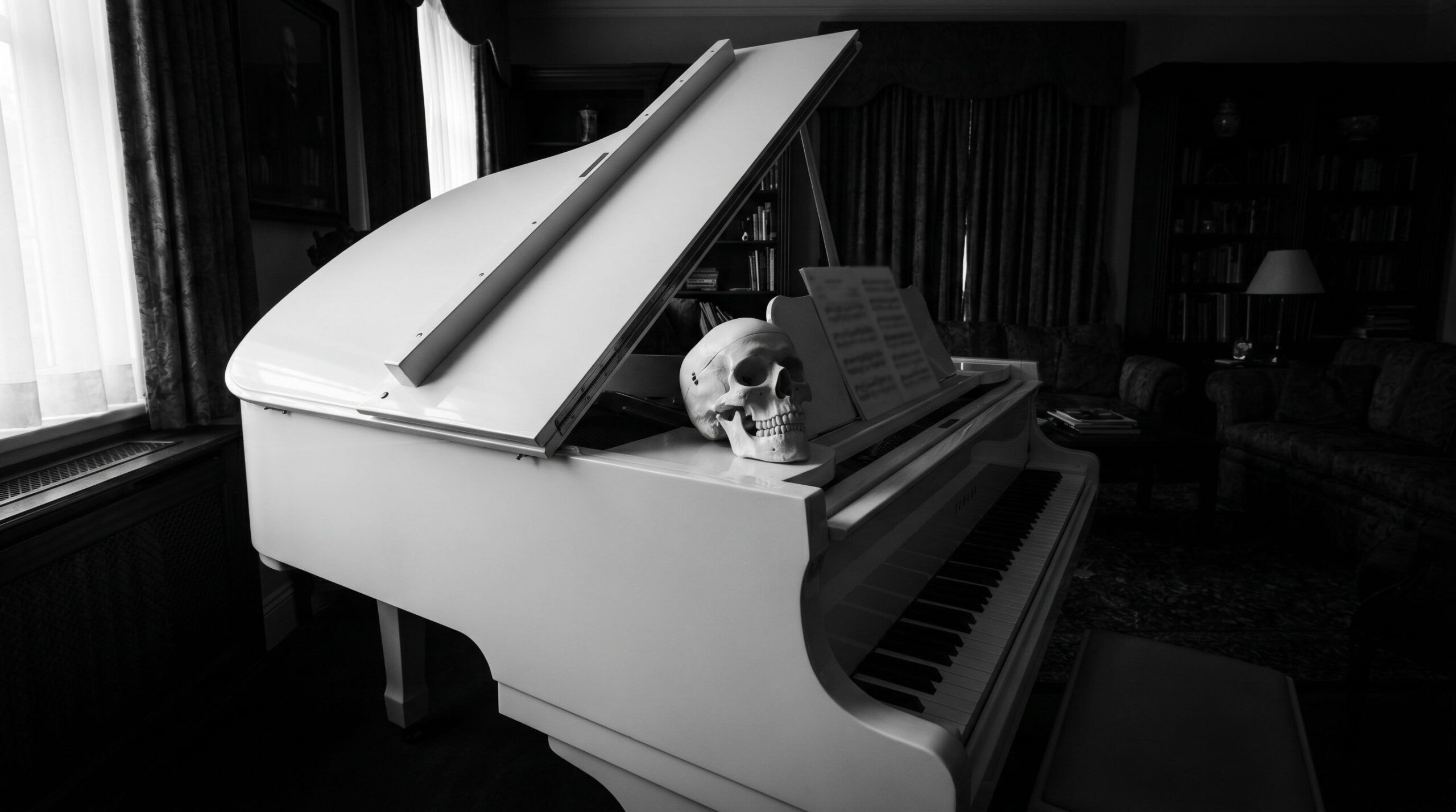
On the drive back from the supermarket, somewhere between the traffic light that is always red and the driveway where I have clipped the same curb for years, the skull on the piano came to mind again. It has stood there for over a year, and I restored it with great care and great affection. The skull of a young woman, recovered during the renovation of a church in the Netherlands. She stands on the piano, in the light, and every time I walk past her I know a little more about her than I did the day before.
I know how that sounds. Most people who hear it pull their shoulders up without meaning to. A dead person's skull in the living room, next to the sheet music, does not fit the picture one has of an evening at home. For me it is the other way around. The longer she stands there, the less she is an object and the more she becomes a person whose story I am piecing together, not out of fantasy, but out of what the bone tells me. And the bone tells a great deal, once you have learned to listen to it.
I have been learning that for a long time, longer than most. I live with skulls, with bones, and I have studied them for over 30 years. I have reassembled them from fragments, when nothing was left of a person but shards. I have restored them properly, when time had worn them down. And I have learned that the moment you touch the bone already decides everything that follows, because a destroyed finding never comes back. So this woman is in good hands, as far as that holds for a person who has been dead for centuries and whose name no one remembers. Whose story would otherwise interest no one.
What I do first, before I ask a single question
Before I begin to read, I see to preservation. That is not a side issue, it is the first duty. A skull that lay in the ground for centuries does not come out intact, it comes out fragile, the surface chalky, the sutures loose, the teeth wobbling in their sockets. Whoever grabs roughly there destroys the very thing they want to understand. So I clean carefully, I stabilize, I secure every single tooth, because no tooth may be lost. A lost tooth is a lost page in a book you cannot open a second time.
Only then do I begin to see. And here I have to admit something that sets me apart from many who see only the bone. I always see the person. After over 15 years of facial reconstruction this is no longer a decision, it is a learned perception that cannot be switched off. I look at a skull and the face begins to lay itself over it, the soft-tissue depths, the shape of the nose, the lips, the gaze. It even runs backward. I see a woman with interesting features in a cafe and I see the skull beneath, the cheekbones, the root of the nose, the angles of the jaw. Creepy, one might think. No, it is the opposite, a gift. It is the deepest form of respect I am capable of, because I never reduce the remains to material. I read them as what they were, a person.
The 4 questions a bone answers long before anyone makes a claim
Forensic and physical anthropology has a framework on which everything else hangs, the biological profile. 4 questions, always the same, always in the same order, and in talks I simply call them the 4 questions. What sex was this person. How old did they get. Where did they come from. How tall were they. From those 4 answers a description forms that turns a nameless heap of bone into a corridor of possibilities, narrow enough that millions suddenly become a handful of missing persons whose files you can lay side by side. Each of these answers rests on methods decades old, calibrated on thousands of known individuals, and each of these methods has limits you must know, otherwise you read things out of the bone it never said. That is exactly what this text is about, what bones give away, and the thin line between reading and inventing.
The man in the next seat who knows everything about skeletons
A few years ago I sat on a flight to Milan, on my way to a training course, a cup of water on the tray table, the belt still loose around my hips. Next to me a man was talking, late 40s, his shirt a touch too tight, with the confidence of someone who had just finished a true-crime podcast. He could not leave, and neither could I, the next hour belonged to us both and the narrow aisle. He was explaining to his young female companion by the window how to read a skeleton. You can see at once whether it is a man or a woman, he said, a professional does that in 2 minutes, and the age too, that is practically written on it. I took a sip of water and said nothing, I smiled to myself and thought of Bones, that series we all loved.
That is the Bones effect, and it is remarkably durable. In the series someone places a bone on a glowing table, a computer beeps, and after 42 minutes including commercials you know the name, the profession and the favorite color of the dead. Reality is slower, more careful and more honest. It is made of probabilities, of ranges, of the constant weighing of several indicators against one another. Whoever claims to read sex in 2 minutes with certainty either got lucky or did not understand the method. Usually the second. The man in the next seat will never notice his error, and that too is a kind of peace.
What the series leaves out is the time. A single skull can occupy me for days, and the best that comes out of it in the end is often a sentence full of qualifications. Probably female, estimated age between 30 and 45, origin in a region with a particular geology, signs of phases of deprivation in childhood. No name, no profession, no favorite color. The layperson is disappointed by sentences like that, because they sound less like a solution than like a reservation. Yet that reservation is the most valuable thing science has to offer. An honest range helps an investigator, an invented certainty leads him astray, and in the worst case it convicts the wrong person. The plane dropped briefly into a gust, the cup trembled on the tray table, and the man in the next seat was just explaining how to read the cause of death off a skull. The bread roll nearly fell from my hand.
Why the pelvis is more honest than the skull
When I want to determine sex, I look at the pelvis first, not the skull. That surprises laypeople, because the skull seems so striking, the brow ridges, the chin, the base of the neck. But the skull deceives more often than it would like to admit. The pelvis does not, because it carries the burden of reproduction, and that burden shaped it long before anyone asked.
The classic method comes from Phenice and dates from 1969, and it is so elegant that it has outlasted half a century. Phenice described 3 traits on the pubic bone, the ventral arc, the subpubic concavity and the medial aspect of the ischiopubic ramus, and he showed that with them sex can be determined with an accuracy of over 95 percent, often from a small fragment (Phenice, 1969). That is the point that makes the method so valuable in practice. You do not need the whole skeleton, a fragment is enough.
Over the decades came the criticism that every good method needs. Klales, Ousley and Vollner presented a revision in 2012 that captures the 3 traits on a 5-step scale and secures them statistically, with a validation of 86.2 percent, and built explicitly to meet the Daubert criteria for admissibility in court (Klales, Ousley & Vollner, 2012). A cleaner, more court-proof version, then. You would think the refinement beats the original.
It does not always. Eliopoulos and colleagues tested both methods in 2023 on the same material, on the Luis Lopes collection in Lisbon, 117 men and 117 women, and the result has a fine, dry irony. The old Phenice method reached 96.5 percent, the modern revision 92.7 percent (Eliopoulos et al., 2023). The original beat its own improvement. Things like that keep science awake. The newest method is not automatically the best, and whoever forgets that mistakes progress for fashion.
When the pelvis is missing, and that happens more often than one would like, the skull remains. It carries a set of traits that differ on the statistical average between the sexes, the nuchal crest, the mastoid process behind the ear, the upper margin of the eye socket, the smooth rise above the root of the nose, the chin region. Male skulls tend toward stronger muscle attachments, more robust ridges, a more angular chin, female ones toward smoother contours and a sharper upper orbital margin. That sounds clear-cut, but it is not, because these traits lie on a continuum, not in neatly separated drawers. There are delicate male skulls and robust female ones, and depending on the population the whole scale shifts. That is why the skull is the second choice. It helps, but it errs more often than the pelvis, and a good finding names the traits one by one and weights them, instead of proclaiming a gut impression.
For the young woman on the piano the matter was clear to my eye, the pelvis was missing, but the cranial traits all ran in the same direction, and nothing contradicted. As sure as a trained eye can ever become without DNA, that is how sure I am with her, and that is not proof, it is a reasoned judgment, something altogether different from a gut feeling. This is the rare, clear case in which all indicators point the same way. The difficult case is the one where they diverge, and then it does not matter which finding shouts loudest, but which is the most reliable.
At this point I have to tell you about a man who taught me how thin that ridge is. Bob, to most people Prof. Robert Mann, one of the most respected forensic anthropologists the field has ever had. It is an honor that I am allowed to call him Bob, and a piece of luck that I was able to learn so much from him. Bob was an anthropologist at the Smithsonian Institution, then for over 20 years at the US military's identification laboratory in Hawaii, the largest skeletal lab in the world, latterly as deputy scientific director, and he founded and led the military's Forensic Science Academy. He has held more than 10,000 skeletons in his hands. He was on the team that identified the remains from the Tomb of the Unknown Soldier. After September 11 he helped identify the victims from the Pentagon, he examined the first victim of the serial killer Jeffrey Dahmer, and in 2023 he traveled to Maui to identify the dead from the fires. He was the first anthropologist in the 200-year history of the College of Physicians of Philadelphia to be named a Fellow. With David Hunt he wrote that photographic atlas of bone disease that stands on my shelf and that I cite further below.
At a training course Bob said a sentence to me that I have never forgotten. Here in Hawaii we have people who donate their bodies to science, he said, and among them are many skulls that according to the records unquestionably belong to women and yet carry distinctly male traits. Since that day I have become more careful. Of course one develops an eye for it over the years, a feeling, I look at a skull and say within seconds, man or woman. But is it also the truth. Can I prove it with court-proof, near-certain confidence. Without DNA, no, and that is something altogether different from guessing. I am one of those people who check themselves again in the middle of their own train of thought and contradict themselves, and that makes me uncomfortable for many around me. I hold up the mirror to everyone, to myself included, and that is not always pleasant. For the woman on the piano all the traits run in the same direction, which is why I am sure. With Bob's Hawaiian skulls they contradict one another, and exactly there, in the contradiction, the error lurks, never in the clear case.
The age is a corridor, not a number
With age it gets delicate, and this is exactly where the man on the plane fails. The age does not stand written on the bone, the age is estimated, and every estimate is a range that widens with the years.
In adults I look at the pubic symphysis, the symphyseal face. Brooks and Suchey delivered in 1990 the system that still sets the standard, 6 phases from I to VI for men and women, calibrated on 1,225 individuals of known age at death from the Los Angeles Medical Examiner's Office (Brooks & Suchey, 1990). A young symphyseal face is rugged, billowing, with pronounced ridges. With the years it smooths, a rim forms, then the breakdown begins. The problem, and Brooks and Suchey pointed it out themselves, is the spread. From phase III on the range becomes so wide that 2 people with the same symphysis can be 20 years apart. Hence the iron rule, never a single indicator alone.
So I take a second. The auricular surface of the ilium, the facies auricularis, described as a method by Lovejoy and colleagues in 1985, changes more regularly across the whole life than the pubic symphysis, which loses its expressive power beyond the third and fourth decade (Lovejoy, Meindl, Pryzbeck & Mensforth, 1985). And I take a third, the cranial sutures. Meindl and Lovejoy showed in 1985 that the lateral-anterior sutures are better age estimators than those of the cranial vault, calibrated on 236 skulls from the Hamann-Todd collection, and they warned in the same breath that suture closure is useful only in combination with other indicators (Meindl & Lovejoy, 1985).
These methods are not all of it. The joints speak along, because their surfaces wear over the years, the smooth covering recedes, bony lips grow at the margins, and a joint that carried load for decades looks different from one that stayed young. The teeth belong here too, their chewing surfaces grind down over a life, and from the degree of this wear one can infer the age roughly, cautiously, because hard fare grinds faster than soft. Above all this, though, stands something that appears in no table, the overall picture. With the years one develops an eye for it, a sum of countless details one no longer names individually. This eye does not replace the method, it guides it, and it is exactly what the man on the plane claimed for himself, without the years behind it. It is clearest with children, because there it is again the teeth that give away the most precise age, and I will come to that shortly.
You see the pattern. Each method on its own is uncertain. Only the bundle yields a defensible corridor. Whoever names a single number is lying, or got lucky. The truth about the age of a skeleton is a range, and an honest range is worth more than an invented precision.
The reason for this uncertainty lies in the biology of aging itself. In a child the body grows on a strict timetable, bone nuclei appear, growth plates close, teeth erupt, all in an order that leaves little room. That is why age estimation in a child is so precise. In an adult growth is finished, and what remains is wear, and wear follows no timetable. It depends on load, on genetics, on illness, on hormones. 2 people of the same age can carry symphyses that seem to lie a decade apart, because one worked hard and the other did not. That is exactly why the range widens with the years. In a 20-year-old I can narrow the age to a few years, in a 60-year-old it becomes a window of 2 decades. That is not a weakness of my work, it is the honest depiction of what the body provides.
For the young woman on the piano the pelvis is missing, and with it the pubic symphysis and the auricular surface, precisely the most reliable age estimators. What remains to me are the cranial sutures, which on their own are the least useful, and the wear of her teeth, which is still slight. Both point in the same direction, an age that is not yet high, and I say no more, because the finding gives no more, and I have learned to stop at the edge of the provable. Where the method ends, fantasy begins, and fantasy has no business at the dissection table.
The body's height hides in the length of a single bone
The fourth question of the profile is the one about stature, and it is often underrated, because it sounds so trivial. How tall was this person. The answer sits in the length of the long tubular bones, above all in the thigh bone, and it follows a surprisingly stable relationship. There is a fixed relationship between the length of the femur and total body height, because the proportions of the body within a population are remarkably reliable. From the measured length of a single complete thigh bone, body height in life can be reconstructed via a regression formula, with an accuracy that is entirely sufficient for narrowing a missing-persons corridor.
The trap lies in the detail, and it is the same one as everywhere in this field. The formulas are population-specific. A regression calibrated on one population estimates wrongly for another, because limb proportions differ between population groups. Whoever applies a European formula to a person of different origin gets a number that looks plausible and is nevertheless wrong. That is why stature hangs on origin, and origin hangs on the enamel, and so in the biological profile one finding reaches into the next, until 4 separate answers become a coherent picture. That is exactly the craft, not the single measurement, but their interplay.
Because this trap has occupied me for years, I built a tool of my own for exactly this calculation, Antrometric, freely accessible and described in detail elsewhere. It takes the measured length of a bone and returns not a bare number, but a documented estimate with everything that belongs to it. The formula family used, the model interval at 90 and 95 percent, the state of preservation of the bone, the agreement of several bones with one another, and an accuracy index that lays open the precision without asserting an identity. You first choose the reference population, otherwise the formula goes wide, then the sex, then the honest state of the bone, that is, no complete bone where only a fragment is present. The estimate comes out at the end as a point, as an interval and as a verdict on reliability, never as a claim about who this person was.
A small thing in it shows how treacherous the field is. A person is taller in the morning than in the evening. The intervertebral discs lose water under the load of the day, and the spine shortens over a waking day by about 1 to 1.2 centimeters. Because the classic reference formulas are calibrated against morning height or against cadaver lengths, a comparison in the afternoon can drag in a small distortion. The tool removes this daily fluctuation on request, a correction for the hours since rising, which over 12 hours adds up to roughly 1 centimeter. So much effort for a few millimeters, and yet it is exactly this effort that separates an honest estimate from a guessed one.
Here too I am helped by the atlas that Robert Mann and David Hunt put together, a regional photographic atlas of bone disease and of normal variation, which grew over years from nearly 10,000 skeletons and which pursues the dry-bone approach as consistently as almost no other work (Mann & Hunt, 2012). It is one of the few books that stand within reach in nearly every osteological lab, and every time you open it it reminds you how thin the ridge is between a pathological finding and normal variation. What looks like disease is sometimes only the span of the normal, and whoever does not know that span sees diseases where there are none.
The smaller skull the universe owed its share to
There is a second skull I have to write about here, and with it the lightness comes harder to me. A child, from England. The change of teeth was almost complete, the permanent teeth almost fully erupted, and it is exactly the teeth that tell its story most precisely, because teeth lie the least of everything a body leaves behind.
In a child age estimation is more precise than in any adult, and the reason lies in dental development. AlQahtani, Hector and Liversidge presented the London Atlas in 2010, an evidence-based atlas that captures age via tooth development and eruption from 28 weeks of gestation to 23 years, calibrated on 72 prenatal and 104 postnatal skeletal remains of known age at death from the collections of the Royal College of Surgeons of England and the Natural History Museum (AlQahtani, Hector & Liversidge, 2010). The material comes from England, and my small skull comes from England, and sometimes a circle closes without your having looked for it. Teeth survive the time in the ground better than almost any bone, and dental age scatters far less than skeletal age, which is why the development of the teeth is the best age estimator a young person leaves us.
On this child's skull I see something that makes me pause every time. Lengthwise grooves in the enamel, linear enamel hypoplasia. Goodman and Rose described in 1990 that these grooves are a non-specific marker of systemic physiological stress in childhood (Goodman & Rose, 1990). Non-specific means they show that there was a phase of need, hunger, illness, deficiency, something disturbed the formation of the enamel. They do not say what it was. They only say that it was. The enamel, in the moment it was forming, stopped growing, and this interruption is frozen, forever.
I write this without pathos, because pathos would be an insult. The child had phases of need, that stands in its teeth, and then it died, before the change of teeth was quite over. The universe stayed in its debt, or the universe owed the debt to the child. I allow myself no more interpretation. The skull stands there, the grooves are there, and my task is to read them, not to embellish them.
Tooth enamel, the black box sealed at 3
Where a person came from is likewise given away by the tooth, and in a way that fascinates me to this day. Tooth enamel is the only tissue in the body that is no longer remodeled after its formation. The bone renews itself throughout life, so it cheats, it overwrites its own past. The enamel cannot do that. What is deposited into it in childhood stays in, unchanged, until the last day.
This makes the enamel a kind of black box, sealed at about 3 years of age and logging the flight thereafter to the end. Strontium is the key. The ratio of strontium isotopes 87 to 86 in the enamel mirrors the geology of the landscape in which a person took up their food as a child, because the strontium travels through plants and water out of the ground into the body and settles in the enamel (Bentley, 2006). The oxygen completes the picture. The oxygen isotopes mirror the water a person drank, and thus the climate of their childhood. Strontium shows above all the geology of the food, oxygen the drinking water, and together they yield a placement, rough but defensible.
Here lies the point many do not grasp. The bone gives away where a person grew up, not where they died. Whoever lived as a child in a region with a particular geology and then moved away carries the first region in the enamel and perhaps the second in the bone, which remodeled itself. So newcomers can be told apart from locals, long after death. The black box blurts out what no passport and no witness can attest anymore.
You have to know the limits of this method, otherwise you overstretch it. Strontium shows no address, it shows a geological type. 2 regions far apart with similar bedrock deliver similar values, and then the enamel alone cannot tell them apart. Only the comparison with local reference values, with the strontium map of a landscape, turns the raw ratio into a statement about origin. And even then it remains a placement within a zone, not a point determination. Whoever claims that an isotope value gives away the home village has not understood the physics. What it gives away is the class of landscape in which a child spent its first years, and that is astonishing enough. For my young woman the enamel points to an origin that fits the region in which she was found, but I say that with all caution, because the finding is a probability, not a stamp.
Something is added that makes the matter lovelier and harder at once. Different teeth form their enamel at different points in childhood. The first permanent molar seals early, a later tooth forms its enamel years afterward. Whoever measures several teeth of the same person reads not a single moment, but a small sequence of childhood, almost like several snapshots from different years. A child that moved between 2 regions can carry that move in its teeth, the early tooth shows the first home, the late one the second. So the black box becomes almost a diary, with few but dated entries.
What a person ate is written in their collagen
Not only origin, diet too writes itself into the body, namely into the collagen of the bone. Schoeninger and DeNiro laid the foundation in 1984. The carbon separates 2 things. It distinguishes plants with different photosynthetic pathways, the so-called C3 from the C4 plants, and it separates terrestrial from marine food sources (Schoeninger & DeNiro, 1984). The nitrogen sharpens the picture. Its value rises with each step of the food chain, about 2 to 3 per mil per trophic level, and it lies on average distinctly higher in animals that ate from the sea than in those that lived off the land.
Concretely that means whoever ate a lot of fish carries a high nitrogen value in the bone, and whoever lived on millet a different carbon value than someone who ate wheat. From 2 isotope values a rough menu forms, not a bill of fare, but a direction. You have to stay careful, a single pair of values can be overstretched, and the clean reconstruction needs several markers. But the principle stands. The dead carry their last years within them as a chemical signature, and whoever reads it knows whether fish often lay on this person's table.
There is one application that touches me especially, because it reaches back into the first months of life. A breastfed child stands one step above its mother in the food chain, because it feeds on her, and this trophic jump shows up as a raised nitrogen value in the tissue formed during the nursing period. When the child is weaned, the value sinks again. From skeletal series one can reconstruct how long a population breastfed and when it weaned, an intimate detail of everyday life that no document ever recorded. The bone remembers the mother's breast, long after both have turned to dust. That is the sort of finding that reminds me I am not working with material, but with the remains of life.
The limit here too, so that no false impression arises. The bone remodels itself, slowly, over years. Its isotope signal is therefore an average of the last years of life, not a photograph of the last day. Whoever wants to know what a person ate in childhood must go back to the tooth enamel, which forgets nothing. Bone and tooth thus tell different chapters of the same life, the tooth the childhood, the bone the last years, and only together do they yield a biography of nutrition.
The petrous bone, the densest vault in the whole body
Now I come to the bone that impresses me most, the petrous bone, the pars petrosa of the temporal bone. It is the densest bone in the human body, and this density makes it a vault. While all other bones lose their DNA over the centuries, the petrous bone holds it fast.
Pinhasi and colleagues showed in 2015 how great this difference is, and the numbers are striking. They examined 10 petrous bones from Holocene contexts in Eurasia, dated between 10,000 and 1,800 years before present, and compared 3 zones within the bone, the spongy part, the dense cortical part around the inner ear, and the densest part within the otic capsule. The endogenous DNA fraction from the densest part exceeded that of the middle zone by up to 65-fold and that of the spongy zone by up to 177-fold (Pinhasi et al., 2015). Gamba and colleagues had earlier shown that petrous samples exceed the DNA yield from teeth by 4 to 16-fold and that from other bones by up to 183-fold (Gamba et al., 2014). The cochlea, the spiral of hearing inside the petrous bone, is therefore today one of the most used elements for recovering human ancient DNA, with its own standardized sampling protocols under clean-room conditions.
Here something interlocks that I will need shortly. The same petrous bone that preserves the DNA over centuries sits exactly in that region of the skull which most often becomes the witness when there is violence. The vault and the scene of the crime lie a few centimeters apart. What the cochlea stores in genetic material can tell us kinship, origin, sometimes even appearance, long after the last witness has died. It opens possibilities no one thought of 20 years ago.
Why the petrous bone preserves so much better than any other bone has to do with its job. It encloses the inner ear, the most delicate sensory apparatus of the body, and nature packed it into the densest bone mass it can build. This density has a side effect that no evolution planned. It protects not only hearing in life, it protects after death the DNA from what otherwise destroys it, from water, from microbes, from the slow chemical decay in the ground. The bone that is remodeled the least and is the densest releases the genetic material most readily. That is a lovely irony of biology, the apparatus for the most fleeting of all senses becomes the most durable store in the whole skeleton.
For practice this means a break with the old approach. In the past, in the search for ancient DNA, one sampled several bones and hoped that one would yield, a poking in the dark with high loss of precious material. Today one goes deliberately to the petrous bone, often to the cochlea within it, and the hit rate rises drastically, while the damage to the skeleton falls. That is the reason this inconspicuous bone, which most people cannot even name, has turned the study of the human past upside down. Whole migrations of prehistory have been reconstructed from petrous bones. And the same bone lies on my piano, in the skull of the young woman from the church.
The tooth I am not allowed to take
There is a question I can answer and that nevertheless plunges me into a crisis of conscience every time, namely the age of the bone itself, that is, how long this person has been dead. For that there is radiocarbon dating, the measurement of the remaining carbon-14 in the tissue. When I have the choice, I prefer a tooth for this measurement. The reason lies in its construction. The tooth is the densest and best-sealed tissue of the whole body, its collagen sits protected inside, shielded against water, against microbes and against the slow exchange with the ground. The porous bone, by contrast, soaks up what the centuries offer it, humic acids from the earth and above all those chemicals with which a find was at some point treated, glues, consolidants, preservatives. Each of these substances drags foreign carbon into the bone and falsifies the measurement, sometimes so thoroughly that even the best cleaning in the lab no longer gets it out, and if a bone glue was even in play, the own can hardly be told from the foreign. What is less contaminated dates more cleanly, and nothing is as well shielded as the collagen deep in the tooth. On top of that a tooth is a clearly delimited sample I can take without touching the rest of the skeleton.
So much for the theory. In practice I have often stood, especially with seized skulls, before a dilemma that no table solves. A fragmented skull is unproblematic, there I take a piece of bone, and the spot is afterward fitted back so professionally that no one ever finds it again. But a skull that belongs to those rare pieces still carrying all 32 teeth in the jaw is something else. To destroy that completeness would be a disgrace, and I cannot bring myself to break a tooth out there or even to detach a piece of bone.
Of course I could replace the extracted tooth optically, with a preparation, a modeled tooth, and I have done that often in the past, so cleanly that no layperson ever notices the difference. But I notice it. A false tooth is not a real tooth. It fills the gap, it deceives the eye, and yet the skull is missing a piece of its truth afterward. That is why, for the valuable pieces, I developed a different solution. Instead of sacrificing a tooth or detaching a visible piece of bone, I drill a fine hole at an inconspicuous spot and take only as much bone mass as the lab really needs, not a crumb more. Then I close the hole with great care and match its color so exactly that the completeness of the skull is preserved and no tooth is missing. For the woman on the piano the question no longer arises anyway. Her teeth stay where they are, every single one.
How violence writes itself into bone
A bone shows not only how a person lived, but often also how they died. A gunshot injury to the skull leaves a signature that can be read, if you know what to look for. The keyword is beveling, the funnel-shaped breakout of the bone along the path of the shot.
The entry wound shows internal beveling, the funnel opens inward, toward the cranial cavity. The exit wound shows external beveling, the funnel opens outward, and it is as a rule larger than the entry wound. From both findings the direction of fire can be reconstructed, even on a heavily shattered skull, if you patiently reassemble the fragments. Radiating and concentric fractures run away from the openings and help to determine the path and the order of several shots.
This reconstruction is handwork, slow and groping. A skull struck by a shot can fall into dozens of pieces, and some of them are so small that an untrained person takes them for worthless and leaves them. Exactly these small pieces often carry the decisive information, the rim of a wound, a piece of beveling, the course of a fracture line. That is why the work does not begin at the table but at the find site, with the careful recovery of every splinter. Whoever is sloppy at the scene cannot rescue at the table what is missing. And when several injuries are present, sometimes their order can be determined, because a later fracture line stops at an earlier one, the way a crack in glass ends at a fissure that was already there. The bone then tells not only that violence occurred, but in what sequence, and that can decide between self-defense and execution.
Just as important is the question of when an injury arose, and here perimortem separates from postmortem. Fresh bone, which still has its organic content, breaks elastically, with irregular edges, because it gives way like green wood. Dry bone breaks brittly, with smooth, straight edges. The SWGANTH guidelines define a perimortem injury as one that carries no signs of healing and at the same time shows no diagnostic traces of postmortem decay, assessed by the biomechanical fracture characteristics of fresh bone (SWGANTH, 2011). Histological research on the microcrack pattern confirms the difference between a fresh and a dry break, but points out honestly that the precise temporal placement remains demanding, especially when a bone is fragmented or still shows fresh properties.
A word of caution belongs here, because perimortem does not mean what the layperson takes it to mean. It denotes the state of fresh bone, not the instant of death. A bone can show perimortem properties although the injury arose hours or even a short time after somatic death, as long as the bone was still fresh. This fine distinction separates forensic anthropology from pathology, and whoever passes over it asserts in court a simultaneity of injury and death that the bone does not provide.
There is one injury pattern that tells its story with particular coldness, the shot to the back of the neck. A single shot into the back of the head or the base of the skull, carried out on a defenseless person. The forensic exhumations of Katyn documented this pattern, the single shot into the base of the skull, the hands bound behind the back, the execution pattern in its full industrial sobriety. I name no weapon model here, because memory alone is not enough as evidence for me, and the finding on the bone carries the passage on its own anyway. The entry at the base of the skull, the internal beveling, the trajectory forward and upward, that is enough to know what happened. The outrage at it stays cold, because the bone does not rage, it reports, and that makes it more unbearable than any adjective.
Disease writes along, and it writes more honestly than any doctor's letter
Diseases leave traces in the bone, and some of them are so characteristic that they allow a diagnosis across centuries. Syphilis is the most striking example. On the outer surface of the skull it can leave caries sicca, a pattern of scarring and destruction that is hard to mistake. And it writes itself into the teeth, when it is congenital.
Congenital syphilis produces Hutchinson incisors, barrel-shaped, narrowed upper incisors notched at the cutting edge, along with the so-called mulberry molars with their knobbly, furrowed chewing surface. Hutchinson described these teeth in works between 1859 and 1888, Moon the molars in 1877, and a re-evaluation of these original works showed that the spectrum was even broader, with notches on lateral and lower incisors, on canine tips, with severe enamel hypoplasia and nodular patterns on the first molars (Ioannou & Henneberg, 2017). The mechanism behind it is cleanly understood. Treponema pallidum, the pathogen, disturbs the ameloblasts during tooth development, that is, the cells that form the enamel, and this disturbance freezes as a malformation in the tooth.
On the cranial vault the same disease can leave caries sicca, a pattern of pits, scars and star-shaped scarring that arises when the inflammation alternately destroys the bone and lets it heal again over a long time. This pattern counts as one of the few that can be assigned with some certainty to treponematosis, in contrast to most other bone changes, which remain ambiguous. Here the real art of paleopathology shows itself, the differential diagnosis. A single finding rarely means anything. Only the coincidence of several findings, the notched teeth with the caries sicca for instance, condenses into a defensible diagnosis. Whoever infers a disease from a single trait errs almost always, because the bone has only a limited number of ways to react to very different ailments.
Here, though, I have to return to the honesty that gives this subject its dignity. Many markers are non-specific. The linear enamel hypoplasia I saw on the child's skull shows stress, not its cause (Goodman & Rose, 1990). Cribra orbitalia, a porous change on the roof of the orbit, and porotic hyperostosis on the cranial vault likewise count as non-specific markers of early metabolic burden, hints of deficiency, infection or illness, without saying which. The older teaching, that they are simply a consequence of iron deficiency, is today considered outdated. This restraint is not a flaw of the method, it is its strength. Whoever attributes to the child a concrete disease the bone does not provide takes its dignity from it. Whoever says honestly, here there was need, its cause I do not know, gives it back.
What the work of a lifetime leaves in bone
The toil of a life also writes itself in, at the attachment sites of the muscles and tendons, the entheses. For a long time these changes were called musculoskeletal stress markers, and it was believed one could read a person's profession from them, the smith from his arm, the carrier from his shoulder. Villotte and colleagues, a whole working group of the discipline, examined such attachment marks on the upper arm in 2010 as markers of load and showed that heavy work does indeed read off the bone (Villotte et al., 2010).
But then came the self-correction, and it is instructive. In 2016 the same research direction deliberately changed the term, from musculoskeletal stress markers to entheseal changes, because physical activity is not the only cause after all, but age, hormones, body build and much more play along, the etiology, then, being multifactorial (Villotte et al., 2016). Heavy work reads off the bone, yes, but not unambiguously, not as a job title. A science that retracts its own term because it promised too much is a grown-up science. Exactly this movement is the mirror into which the layperson in the next seat never looks.
An example makes the trap vivid. A strongly developed attachment on the upper arm can come from years of lifting heavy loads. But it can also simply reflect age, because these changes increase with the years anyway, or a genetic predisposition, or an inflammation that was gone through. 4 people with the same finding can have lived 4 entirely different lives. Whoever turns the strong attachment into the smith tells a story that sounds good and can be wrong. More serious is the cautious statement, this person loaded their arm strongly over a long time, the kind of load I do not know. That is less cinema, but it is true, and truth in my profession is worth more than a good story.
A warning to the reader, with the usual pinch of polemic
Now I have to raise my voice against my own guild, and against all who want to read bones without having learned their language. The bone does not lie. But it is lied about, constantly, by whoever reads more out of it than is in it. That is the real danger, not the honest doubt of the method, but the dishonest certainty of the one who presses the range into a number, the non-specific marker into a diagnosis, the attachment mark into a profession.
The Otto Sapiens in the next seat on the plane is the harmless version, he impresses only his companion. The dangerous version sometimes sits in the same field as I do, it wears a title and a certainty the bone never covered. It tells a chamber, a court, a public, that the finding is unambiguous, where it is not. Meindl and Lovejoy warned, never a single indicator alone, and the entheses researchers defused their own term because it claimed too much. Whoever ignores these warnings no longer reads, he invents. And an invented bone is more dangerous than none at all, because it wears the garment of science without its humility.
There is a reason this troubles me more than the vanity in the next seat. Over-interpretation has consequences. An age proclaimed too confidently, a marker interpreted too unambiguously, an invented job biography, all of that can wander into a file, into an expert report, into a verdict. That concerns not only my own guild. It concerns also those archaeologists who have learned for a few months how one supposedly reads bones correctly, and afterward trust themselves to read a whole life out of one finding. What sometimes comes under my eyes from such reports makes my hair stand on end, not out of vanity, but because a confidently misread bone does the same damage as a false witness statement, except that no one contradicts it. The dead cannot defend themselves, and the living, whom a false finding burdens, often cannot either. The humility of the method is therefore no academic coquetry, it is an ethical duty. I would rather say 3 times, the bone does not provide that, than once assert a certainty that exists only in my head. The science that knows and states its own limits is not the weaker one, it is the only one you may trust. Everything else is the Otto Sapiens with a doctorate, and he is more dangerous than his brother on the plane, because someone believes him.
What remains when the face has gone
She still stands on the piano, the young woman from the church, and as I write this I know more about her than on the day she came to me. I know roughly how old she got. I know where the enamel of her teeth comes from. I know the traces that life wrote into her. I have given her face back to her with love and the fullest devotion. And I know what I do not know, and that is just as important.
A question remains, and it has nothing to do with method. What happens to her when I am no longer here. Who will read her then, who will preserve her, who will see in her the person and not the object. To that I have no answer, only the duty to treat her as a person for as long as I can.
Sometimes, when I walk past the piano in the evening and the light from the street falls on the skull, I see not the bone but her face. I know it, I see it every day, because I gave it to her, the cheeks over the cheekbones, the nose over the aperture, the gaze over the sockets. I reconstructed her face, the way I have reconstructed the face of many of my skulls, because that is how I learned this craft, layer by layer the soft tissue over the bone, until a person looks back whom no one has seen for centuries. She was young. She came from somewhere her enamel still knows. She had a life of which her teeth and her sutures have kept a few pages. The rest I owe her, and part of that debt is not to over-read her, not to ascribe to her what is not in her.
She stands in the light, on the piano, and looks at me.
References
- AlQahtani, S. J., Hector, M. P., & Liversidge, H. M. (2010). Brief communication: The London atlas of human tooth development and eruption. American Journal of Physical Anthropology, 142(3), 481-490. https://doi.org/10.1002/ajpa.21258
- Bentley, R. A. (2006). Strontium isotopes from the earth to the archaeological skeleton: A review. Journal of Archaeological Method and Theory, 13(3), 135-187.
- Brooks, S., & Suchey, J. M. (1990). Skeletal age determination based on the os pubis: A comparison of the Acsádi-Nemeskéri and Suchey-Brooks methods. Human Evolution, 5, 227-238.
- Eliopoulos, C., et al. (2023). Sex assessment from the pelvis: A test of the Phenice (1969) and Klales et al. (2012) methods. Forensic Science, Medicine, and Pathology, 20(3), 778-786. https://doi.org/10.1007/s12024-023-00685-4
- Gamba, C., Jones, E. R., Teasdale, M. D., McLaughlin, R. L., Gonzalez-Fortes, G., Mattiangeli, V., et al. (2014). Genome flux and stasis in a five millennium transect of European prehistory. Nature Communications, 5, 5257. https://doi.org/10.1038/ncomms6257
- Goodman, A. H., & Rose, J. C. (1990). Assessment of systemic physiological perturbations from dental enamel hypoplasias and associated histological structures. Yearbook of Physical Anthropology, 33, 59-110.
- Ioannou, S., & Henneberg, M. (2017). Dental signs attributed to congenital syphilis and its treatments in the Hamann-Todd Skeletal Collection. Anthropological Review, 80(4), 449-465. https://doi.org/10.1515/anre-2017-0032
- Klales, A. R., Ousley, S. D., & Vollner, J. M. (2012). A revised method of sexing the human innominate using Phenice's nonmetric traits and statistical methods. American Journal of Physical Anthropology, 149(1), 104-114. https://doi.org/10.1002/ajpa.22102
- Lovejoy, C. O., Meindl, R. S., Pryzbeck, T. R., & Mensforth, R. P. (1985). Chronological metamorphosis of the auricular surface of the ilium: A new method for the determination of adult skeletal age at death. American Journal of Physical Anthropology, 68(1), 15-28.
- Mann, R. W., & Hunt, D. R. (2012). Photographic regional atlas of bone disease: A guide to pathologic and normal variation in the human skeleton (3rd ed.). Charles C. Thomas.
- Meindl, R. S., & Lovejoy, C. O. (1985). Ectocranial suture closure: A revised method for the determination of skeletal age at death based on the lateral-anterior sutures. American Journal of Physical Anthropology, 68(1), 57-66. https://doi.org/10.1002/ajpa.1330680106
- Phenice, T. W. (1969). A newly developed visual method of sexing the os pubis. American Journal of Physical Anthropology, 30(2), 297-301. https://doi.org/10.1002/ajpa.1330300214
- Pinhasi, R., Fernandes, D., Sirak, K., Novak, M., Connell, S., Alpaslan-Roodenberg, S., et al. (2015). Optimal ancient DNA yields from the inner ear part of the human petrous bone. PLOS ONE, 10(6), e0129102. https://doi.org/10.1371/journal.pone.0129102
- Schoeninger, M. J., & DeNiro, M. J. (1984). Nitrogen and carbon isotopic composition of bone collagen from marine and terrestrial animals. Geochimica et Cosmochimica Acta, 48(4), 625-639.
- Scientific Working Group for Forensic Anthropology (SWGANTH). (2011). Trauma analysis (Revision 0). https://www.nist.gov/system/files/documents/2018/03/13/swganth_trauma.pdf
- Villotte, S., Castex, D., Couallier, V., Dutour, O., Knüsel, C. J., & Henry-Gambier, D. (2010). Enthesopathies as occupational stress markers: Evidence from the upper limb. American Journal of Physical Anthropology, 142(2), 224-234. https://doi.org/10.1002/ajpa.21217
- Villotte, S., Assis, S., Cardoso, F. A., Henderson, C. Y., Mariotti, V., Milella, M., et al. (2016). In search of consensus: Terminology for entheseal changes (EC). International Journal of Paleopathology, 13, 49-55. https://doi.org/10.1016/j.ijpp.2016.01.003

Is It Legal to Own a Real Human Skull? Yes, Subject to 3 Conditions
Private possession of a real human skull is not prohibited in Germany. Three conditions decide the question, and…

The Science and Ethics of Age-Gap Attraction: Why Older Men Are Drawn to Younger Women, and Where the Line to Crime Runs
A forensic examiner lays out the evolutionary psychology and the data from 130 countries: the algorithm of age-gap…
The Pressure Fallacy: What Pink Teeth in the Dead Really Reveal, and What They Do Not
A forensic investigation through 200 years of literature, prompted by three cases, two of which I examined myself,…