The Architecture of Becoming: What a Child’s Skull Reveals About Biological Precision and Its Paradox

On the 7-year developmental program that produces a permanent dentition, what forensic odontologists read from enamel and root formation in mixed dentition specimens, and the particular discomfort of studying the biological intricacy of a species whose history is also a record of its willingness to destroy the children it produces.
The skull is prepared in the manner that made it famous as a teaching specimen, though the word “famous” sits awkwardly in this context. The osseous structures on the left side of the face have been meticulously removed by a skilled preparator, a technique that reached its highest expression in the anatomical workshops of the 18th and 19th centuries and that requires both surgical precision and a detailed understanding of what lies beneath what you are cutting away. The preparation exposes the dental architecture of both the maxilla and the mandible simultaneously: a complete view of 2 timelines running in parallel, the deciduous teeth still in place in their sockets, the permanent successors visible beneath them in various stages of formation, their crowns already shaped, their roots still growing toward the eruption sequence that will begin within the next 1 to 3 years.
This is a child of approximately 6 to 8 years. The specimen is part of the London reference collection, which forms one of the empirical foundations of the London Atlas of Human Tooth Development and Eruption, published in 2016, and which represents one of the most precise currently available tools for the estimation of chronological age from dental morphology in children and adolescents (Adams et al., 2018, “Impact of population-specific dental development on age estimation using dental atlases”, American Journal of Physical Anthropology, 168(1), 190-199). Looking at the prepared skull, you see a biological program in mid-execution, precisely where it was designed to be at this particular moment in a child’s development, unaware of and indifferent to whatever happened afterward.

The Anatomy Tradition Behind the Preparation
The preparation technique applied to this skull has a history that is worth a brief acknowledgment before discussing its scientific content. The systematic removal of overlying bone to expose underlying dental structures was practiced by anatomists from at least the 17th century, initially as a teaching device for medical students who needed to understand the relationship between the visible oral cavity and the developmental architecture beneath it. The preparation of pediatric skulls for dental education was particularly refined in the 19th century, when the explosion of dental schools across Europe and North America created a demand for demonstrable specimens that illustrated the normal developmental sequence to cohorts of students who could not otherwise examine it in the living patient.
The best of these preparations achieved what this specimen achieves: a complete exposure of both the deciduous and permanent dentitions in a single plane of view, allowing the observer to see simultaneously what is visible in the mouth of a living child and what lies beneath the visible surface, ready to emerge in the sequence that will play out over the next 5 to 7 years. The skill required is not trivial. The cortical bone of a child’s maxilla and mandible is thinner than adult bone, and the relationship between the overlying structures and the underlying tooth follicles is close, meaning that an imprecise cut removes evidence rather than exposing it. The specimens that survive from major collections survived because they were made well.
Odontogenesis: The Program That Begins Before the World Begins for the Child
Tooth formation begins during the sixth week of embryonic development, before the human fetus has a face in any form a non-specialist would recognize. The dental lamina, a thickened band of oral epithelium, invaginates into the underlying mesenchyme along the future dental arches and initiates the sequence of bud formation, cap stage, bell stage, and matrix deposition that will ultimately produce the deciduous dentition. This process, odontogenesis, proceeds with a genetic choreography that has been conserved across mammalian evolution and that is, for the purposes of forensic age estimation, one of the most stable and therefore most reliable biological timekeepers available in the human body.
The deciduous dentition, 20 teeth in total, erupts over a period beginning at approximately 6 months after birth and concluding by approximately 30 months, following a sequence that begins with the lower central incisors and concludes with the second molars. These teeth are not placeholders in any superficial sense; they perform the full mechanical and physiological functions of dentition throughout early childhood, they are morphologically distinct from their permanent successors in ways that inform both clinical management and archaeological identification, and they are, in the skull before us, still in their sockets, still intact, still carrying the wear patterns and enamel morphology that document the dietary history of the first 6 to 8 years of this individual’s life.
The permanent dentition begins its formation before the deciduous teeth have finished erupting. The crown of the first permanent molar, the tooth whose eruption at approximately 6 years is the most significant single landmark in pediatric dental development, begins forming during the third trimester of fetal life, its occlusal surface depositing enamel prisms while the child is still in utero, its roots extending and completing in the years following its eruption around the 10th year. The first permanent molar is the cornerstone of the permanent arch, and its eruption marks the beginning of the mixed dentition period, that approximately 6-year interval during which deciduous and permanent teeth coexist in the oral cavity in a continuously shifting ratio, with the deciduous roots resorbing under the chemical pressure of the erupting successors and the permanent crowns emerging through the alveolar bone in a sequence that is partly genetically programmed and partly modified by local mechanical and nutritional conditions.
What Mixed Dentition Looks Like: The Skull at Six to Eight Years

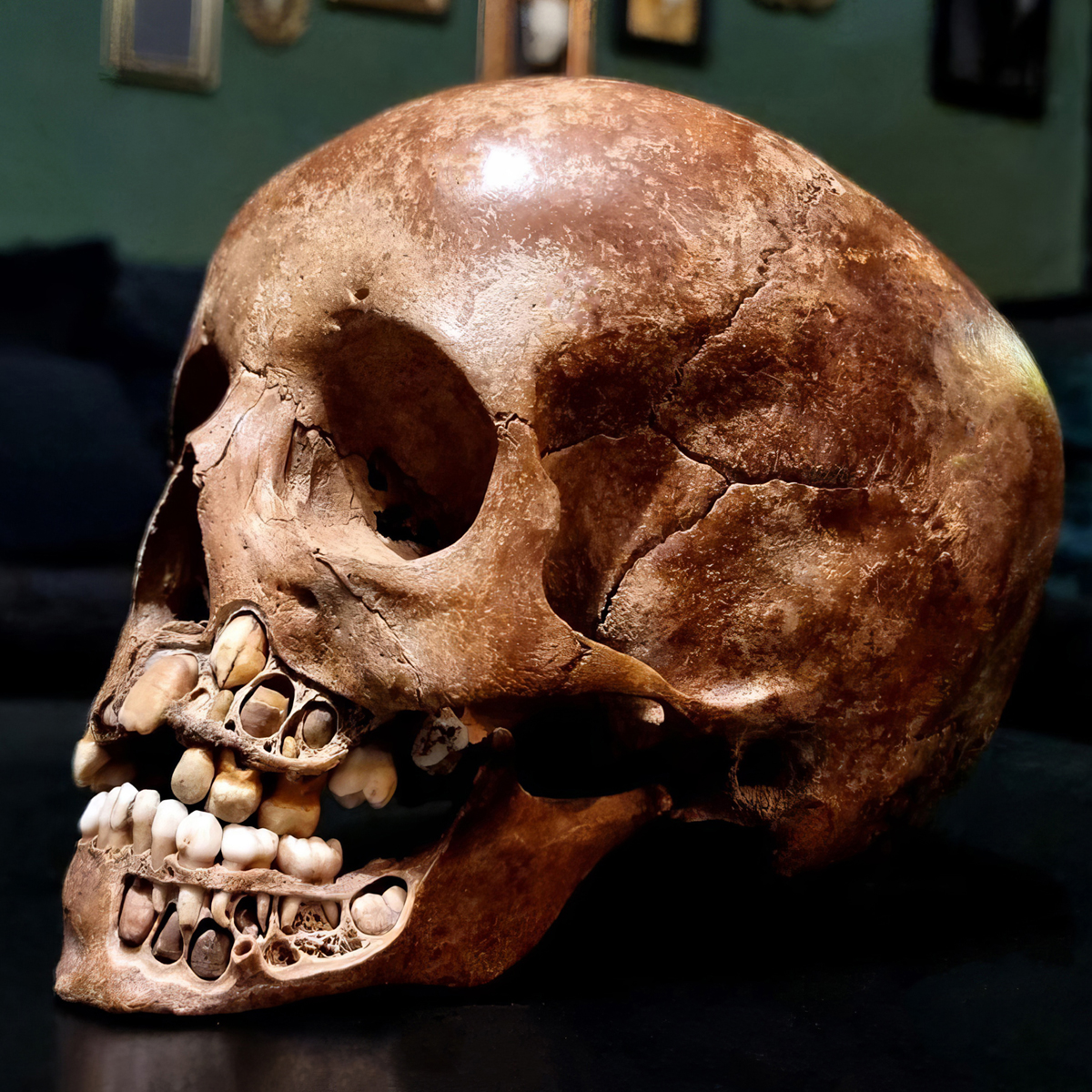

The prepared skull in the London reference collection is a forensic and educational document of considerable precision. In a child of 6 to 8 years, the dental status is broadly predictable: the lower central incisors have typically already erupted in permanent form, the upper central incisors may be in transition, the first permanent molars have erupted in all 4 quadrants. Beneath the alveolar bone and the deciduous canines and premolars, the permanent successors are visible in various stages of root elongation, their crowns complete, their apices open, their timeline written in the relative proportions of crown to root that form the basis of the most precise forensic age estimation methods currently in use.
The permanent lateral incisors, canines, premolars, and second molars are all present in various stages of development in this age range, each at a stage that the skilled analyst can map against reference populations to produce an age estimate. The combination of stages across multiple teeth, each developing on a slightly different schedule, provides a multipoint temporal estimate whose precision exceeds that of any single tooth taken in isolation. This is the fundamental logic of multi-tooth age estimation methods: no individual tooth is a sufficiently precise clock, but 7 developing mandibular teeth observed simultaneously produce an age estimate whose error margin, under favorable conditions, falls within approximately plus or minus 1 year.
Forensic Age Estimation: Demirjian, London Atlas, and the State of the Evidence
The systematic estimation of age from dental development in children is an area of forensic odontology with a substantial methodological literature, a genuine and productive scientific debate, and a practical significance that extends from the identification of unknown remains to the determination of the legal status of asylum-seeking minors whose documentary records are either absent or contested.
Demirjian and colleagues published the foundational method in 1973, based on a 7-stage system applied to the mandibular teeth of French-Canadian children, in which each tooth is assigned a developmental stage from A (initial cusp formation) through G (root apex closed), and the combination of stages across 7 teeth is converted to an age estimate through population-specific weighting tables (Demirjian, A., Goldstein, H., & Tanner, J.M., 1973, “A new system of dental age assessment”, Human Biology, 45(2), 211-227). The method has been applied to populations worldwide with variable results: a meta-analysis of 26 studies covering 11,499 children found that Demirjian’s method overestimated chronological age by an average of 0.35 years in males and 0.39 years in females, with the magnitude of overestimation varying substantially by population (Li et al., 2012, “Assessment of dental age of children aged 3.5 to 16.9 years using Demirjian’s method”, PLoS ONE, 7(12), e51601).
The London Atlas of Human Tooth Development and Eruption, published in 2016 and based on a large sample of British children with known birth dates, provides a visual reference standard against which the observed dental status of an unknown individual can be compared directly, without the mathematical weighting of the Demirjian method (Cameron, N., et al., 2016, London Atlas). Recent comparative studies have found that the London Atlas outperforms Demirjian’s method in precision for several populations: in a 2024 study of 711 Saudi children, the London Atlas produced mean differences from chronological age of 0.03 years for males and 0.00 years for females, compared to 0.55 and 0.76 years respectively for the Demirjian method (PMC11394459, 2024). The London Atlas’s greater accuracy appears to result from its more granular staging system and its accommodation of individual tooth variation, whereas Demirjian’s fixed weighting system introduces systematic bias when applied to populations whose dental maturation rates differ from the French-Canadian reference sample.
Current best practice in forensic odontology for age estimation in children combines multiple methods, typically the London Atlas alongside Willems’ modification of the Demirjian system, and reports the result as an age range with an explicit confidence level rather than as a point estimate. A child whose dental development corresponds to the London Atlas standard for 7.5 years is estimated to be between 6.5 and 8.5 years of age at the 95 percent confidence level, which is operationally useful for triage in missing persons investigations and for the legal determination of minority status in asylum proceedings, while remaining honest about the limits of the measurement.
Forensic Applications Beyond the Laboratory: Age Determination in the Field
The practical significance of dental age estimation in forensic contexts extends considerably beyond the examination of remains in a controlled laboratory setting. 3 specific application areas deserve particular attention because they illustrate the range of contexts in which the methods described above are operationally critical.
The first is the identification of unknown human remains from mass casualty events, whether natural disasters, transportation accidents, or deliberate acts of violence. In these contexts, the remains of individuals without documentation or prior dental records may require age determination from dental development to enable probabilistic identification against missing persons databases. Children present particular challenges in this context because their relatively rapid developmental change means that the passage of even 6 months between a missing persons filing and a recovery may produce discernible changes in the dental stage, requiring the analyst to apply appropriate developmental timelines to the interval rather than treating the match as static.
The second application is the legal determination of age in unaccompanied asylum-seeking minors. European jurisdictions, including Germany, France, the Netherlands, and the United Kingdom, have established legal frameworks for the forensic assessment of age in individuals who claim minority status but whose documentary evidence is either absent, untraceable, or disputed. Dental age estimation using the London Atlas and related methods, combined with skeletal maturation assessment of the wrist and hand, is a standard component of these assessments in several jurisdictions. The ethical and legal dimensions of this application are extensively debated, with the Council of Europe and several national pediatric and odontological societies having issued position statements addressing the appropriate use and limitations of the methods, the minimum standards for reporting, and the legal consequences of different levels of diagnostic certainty (Council of Europe, 2017, “Children deprived of liberty and unaccompanied asylum-seeking children”).
The third application, and the one most directly connected to the specimen documented here, is the development and validation of population-specific reference standards. The London reference collection is an identified collection, meaning the age at death, sex, and origin of each individual are documented, which allows the specimens to serve as calibration material for age estimation methods applied to unidentified individuals from similar populations. The value of identified reference collections to forensic anthropology cannot be overstated: without them, the accuracy claims of age estimation methods are essentially claims about the relationship between developmental staging and chronological age that cannot be independently verified. With them, the error distribution can be computed, the systematic biases identified, and the confidence intervals stated with empirical grounding rather than theoretical assumptions.
What Teeth Tell Us Beyond Age
The developmental record encoded in a child’s dentition extends considerably beyond age estimation, in directions that are both clinically and historically significant.
Enamel hypoplasia, visible as linear defects in the enamel surface running parallel to the incremental lines of growth, marks periods of metabolic stress during crown formation, whether from nutritional deficiency, febrile illness, or other systemic disruption. Because the timing of enamel formation is known with reasonable precision for each tooth and each region of each crown, the location of a hypoplastic defect on the crown surface can be dated to the approximate age at which the stress occurred. A defect at the cervical margin of the upper central incisor crown places the stress event at approximately 3 to 4 years of age, when that region of the crown was forming. Multiple defects on multiple teeth allow the reconstruction of a stress timeline covering the first decade of life, which is what makes enamel hypoplasia such a useful tool in both the forensic examination of individual remains and in the population-level study of health and nutrition in past societies.
Dental caries prevalence and pattern document dietary composition, particularly sugar exposure frequency and carbohydrate consistency, in a way that skeletal measurements cannot. The introduction of refined carbohydrates into a population’s diet typically produces a rapid and visible increase in caries prevalence that is archaeologically traceable in skeletal samples from successive time periods. This is one of the reasons that dental analysis forms a standard component of bioarchaeological site reports: the teeth carry a dietary history that the rest of the skeleton largely does not.
Third molar agenesis, present in approximately 20 to 25 percent of European populations, reflects both genetic factors and the ongoing evolutionary trajectory of jaw size reduction in anatomically modern humans. The third molar is, in a functional sense, becoming vestigial across portions of the global population, a process driven by the dietary and cooking practices of Homo sapiens over the last 10,000 years, which have reduced the mechanical demands on the posterior dentition to the point where the genetic program for third molar formation is no longer under strong positive selection in all human populations. This represents one of the more visible examples of recent microevolutionary change in the human body, and it is visible in the teeth.
The cusp morphology of the permanent teeth, their specific fissure patterns, cusp numbers, and developmental groove configurations, carries population-specific genetic information that has been used for forensic identification purposes and for the reconstruction of prehistoric migration patterns. Shovel-shaped incisors, for example, are found at high frequency in East Asian and Indigenous American populations and at low frequency in European and sub-Saharan African populations, a distribution that reflects the microevolutionary history of each group and that provides supplementary population affinity information in forensic cases where the skeletal sample is otherwise ambiguous.
The prepared skull in these photographs is, in this sense, not a specimen but a document, one written in a language that requires years of training to read and that rewards patient study with a degree of biographical information that no external examination of the skull surface could approach.
The Paradox That Refuses to Dissolve
There is a particular quality to the experience of working with child remains, whether ancient or modern, whether archaeological or forensic, that I have never been able to separate from the scientific work itself. The biological precision on display in the mixed dentition of a 6- to 8-year-old skull, the genetic choreography of the developmental program, the accuracy of the timing, the resilience of the enamel, the sequencing of eruption that has been conserved across thousands of generations, represents an order of engineering that is genuinely remarkable regardless of one’s views about its ultimate origin. The human genome contains the instructions to build this structure, and those instructions execute correctly under an enormous range of environmental conditions, with a fidelity that materials scientists would describe as extraordinary.
The same species whose genome contains those instructions has produced a documentary record of its treatment of children that is, to put it in the mildest available terms, inconsistent with the biological investment represented by the developmental program. The archaeological record is continuous on this point across cultures and millennia, and the contemporary record continues it without interruption. The child whose skull is documented here was not harmed by human action. But the research tradition in which this specimen participates exists partly because forensic anthropologists need reliable methods for determining the age of the young remains they encounter in circumstances where the question of how those remains came to be where they are is a matter of criminal investigation.
The precision in age estimation that methods like the London Atlas and Demirjian’s system achieve serves, among other functions, the documentation of violence against children in terms that are legally usable, scientifically defensible, and capable of surviving cross-examination in court. That these methods are needed, that the forensic literature on pediatric age determination is extensive and actively developing, is itself a statement about the world in which they operate. I do not draw comfort from the sophistication of the methodology.
The Antrometric platform at antrometric.com applies quantitative age estimation from skeletal and dental measurements with the precision that current reference standards support, refined as population-specific data accumulates across the institutions and cases to which it is applied. It does not resolve the paradox. Nothing does. But precision in documenting the remains of children is, at minimum, a contribution to the possibility of accurate documentation, which is the precondition for anything that might eventually follow.
Closing
The 6-year molar erupts on schedule regardless of what is happening around the child who carries it. Its roots complete on schedule regardless of whether the environment in which that completion occurs is adequate to support the rest of what the child requires. The developmental program is not aware of its context. It executes, precisely and without sentiment, the instructions encoded in the genome of a species that has been, over the span of its existence, simultaneously the author of this program and the most significant threat to the individuals in whom it runs.
I hold both of those things clearly when I look at this skull, and I have not yet found a framework that makes holding them simultaneously comfortable. I do not expect to find one.
References
- Adams, D.M., Ralston, C.E., Sussman, R.A., et al. (2018). Impact of population-specific dental development on age estimation using dental atlases. American Journal of Physical Anthropology, 168(1), 190-199.
- Avery, J.K., & Ancowitz, A.N. Oral Development and Histology. Thieme.
- Cameron, N., et al. (2016). London Atlas of Human Tooth Development and Eruption. University College London.
- Cameron, A.C., & Widmer, R.P. Handbook of Pediatric Dentistry. Elsevier Mosby.
- Chowdhry, A., Kapoor, P., Bhargava, D., Bagga, D.K., & Mehta, A. (2023). Comparison of Demirjian’s comprehensive chart with the London atlas of tooth development in children and adolescents: A pilot study. Forensic Sciences Research, 8(4), 332-339.
- Demirjian, A., Goldstein, H., & Tanner, J.M. (1973). A new system of dental age assessment. Human Biology, 45(2), 211-227.
- Larsen, W.J. Human Embryology. Churchill Livingstone.
- Li, G., et al. (2012). Assessment of dental age of children aged 3.5 to 16.9 years using Demirjian’s method: A meta-analysis based on 26 studies. PLoS ONE, 7(12), e51601. https://doi.org/10.1371/journal.pone.0051601
- PMC11394459. (2024). A comparison of two methods of dental age estimation in a population of Saudi children and adolescents. International Journal of Environmental Research and Public Health, MDPI.
- Scott, G.R. The Anthropology of Modern Human Teeth. Cambridge University Press.

Is It Legal to Own a Real Human Skull? Yes, Subject to 3 Conditions
Private possession of a real human skull is not prohibited in Germany. Three conditions decide the question, and…

The Science and Ethics of Age-Gap Attraction: Why Older Men Are Drawn to Younger Women, and Where the Line to Crime Runs
A forensic examiner lays out the evolutionary psychology and the data from 130 countries: the algorithm of age-gap…
The Pressure Fallacy: What Pink Teeth in the Dead Really Reveal, and What They Do Not
A forensic investigation through 200 years of literature, prompted by three cases, two of which I examined myself,…