Corpse Stains: Formation, Persistence and Significance in Forensic Photography
Corpse stains, also known as livores, form a complex mosaic of discolorations that has left the forensic world in awe for centuries. These postmortem markings, which occur as blood accumulates in the lowest parts of the body after death, offer a treasure trove of information invaluable for determining the time of death and conducting extensive forensic investigations. But what exactly lies behind these enigmatic stains that appear like silent witnesses after a person’s passing?
With the final heartbeat, not only does life end, but also the circulation of blood ceases, allowing it to follow the laws of gravity and settle in the deeper body regions. This accumulation results in the characteristic dark violet to bluish discolorations known as corpse stains. Within just a few hours after death, these stains become visible, and their detailed examination can provide forensic experts with valuable clues about the circumstances surrounding the death. It is more than a simple result of gravity: the way the blood disperses and the patterns that emerge can shed light on the body’s position at the time of death and any subsequent changes.
Modern forensic science has made remarkable progress over recent decades, particularly through the use of state-of-the-art imaging technologies and spectral analyses. These innovations allow corpse stains to be documented and analyzed with a precision that was once simply unimaginable. By employing UV light and infrared cameras, changes in tissue that remain hidden to the naked eye become visible. This technological revolution has significantly expanded the forensic toolbox and increased the accuracy of investigative procedures.
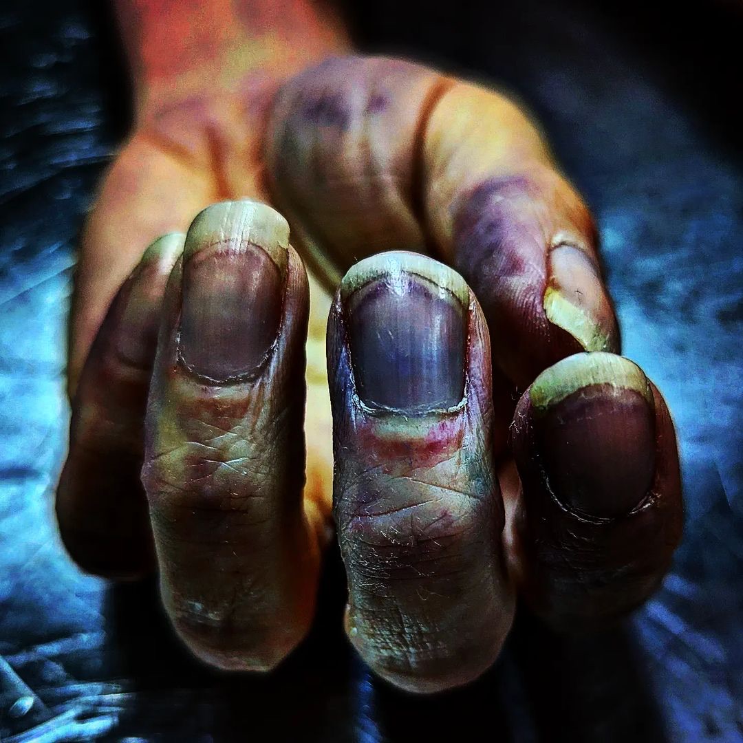
The color changes, distribution and intensity of corpse stains can provide clues to the cause of death and the postmortem process. While differences in coloration may indicate poisoning, atypical patterns often suggest physical trauma or a change in the body’s position after death. A particularly fascinating aspect is the study of the molecular changes that occur postmortem and influence the formation of corpse stains. With the latest biochemical and genetic analysis methods, this process is becoming increasingly understood, not only improving the estimation of time of death but also expanding our knowledge of the dying process and postmortem decay.
Almost immediately after death, the intricate spectacle of corpse stain formation begins. Within 20 minutes to three hours, initial changes appear, though this process is fluid and exhibits various phases that offer valuable information for forensic analysis. In the initial phase, known as the hypostatic phase, the stains are not yet fixed and can shift if the body is moved. This phase lasts for up to about six hours, during which gravity may cause the blood to flow into different body regions if the body is repositioned.
Between six and twelve hours after death, the post-hypostatic phase sets in, during which the corpse stains begin to fix. This fixation results from the leakage of blood plasma into the surrounding tissue and the simultaneous coagulation of the blood within the vessels. This is a critical indicator for forensic examination, as it sheds light on the postmortem progression and potential tampering with the body. The final fixation of corpse stains occurs after roughly twelve hours and is usually complete after 24 hours, making the stains immovable regardless of any subsequent body movements.
The factors influencing the formation and distribution of corpse stains are diverse. The condition of the blood vessels, the composition of the blood and individual physiological differences of the deceased all play a role, as do external factors such as temperature and humidity. Interestingly, pathological conditions such as blood diseases or poisonings can also alter the appearance of corpse stains. For instance, certain toxins like carbon monoxide can tint the stains a bright pink, providing clear evidence of poisoning.
The analysis of corpse stains engages a fascinating and technically demanding branch of science that requires in-depth knowledge of physiology and pathology. Modern imaging techniques such as spectral photography have greatly expanded the possibilities for documenting and examining these stains. These technologies allow subtle tissue changes to be visualized that remain imperceptible to the naked eye. Their significance is underscored by their ability to capture detailed and precise information about the circumstances of death and postmortem changes.
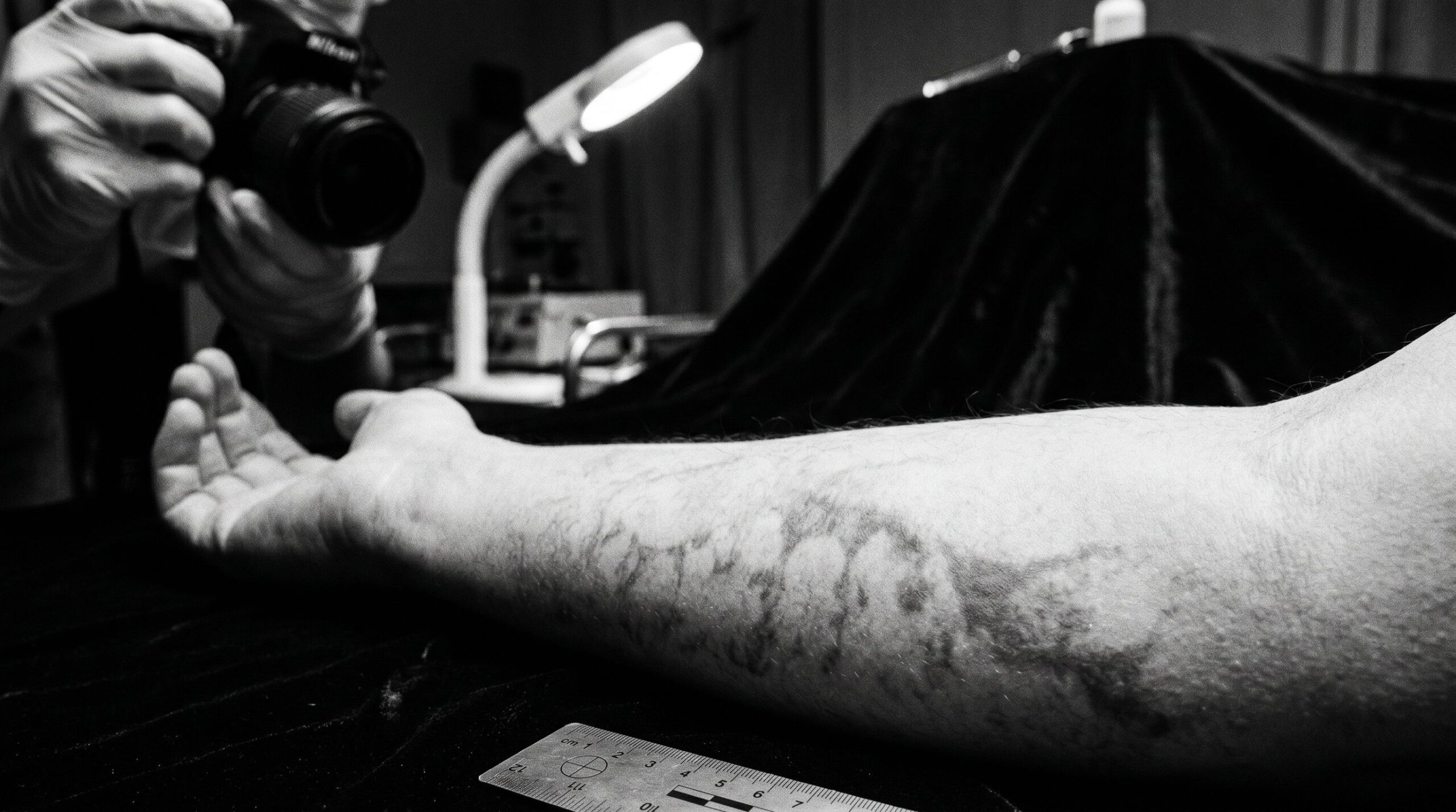
Traditionally, documentation of corpse stains begins at the scene with a visual inspection by a detective. This initial observation is crucial because it provides clues about the body’s position at the time of death and any subsequent alterations. The advent of forensic photography made it possible to create high-resolution images that serve as objective and permanent evidence. Spectral imaging goes a step further by utilizing various wavelengths of light to highlight details not visible in the standard spectrum, leading to even more precise documentation and analysis of corpse stains.
The digitization and automation of data analysis have revolutionized forensic documentation. Cutting-edge image processing software now enables the analysis and comparison of captured images, resulting in a more precise determination of stain patterns and their changes over time. These digital tools offer greater accuracy and objectivity, which is especially important when presenting evidence in court.
A notable case is that of Jeffrey Epstein, where the precise analysis of corpse stains, conducted by Dr. Michael Baden, provided crucial clues indicating possible external interference. This work demonstrated how vital the accurate investigation and documentation of corpse stains can be for solving cases and ensuring justice.
In summary, corpse stains are far more than a sign of postmortem change. They are an essential tool in forensic investigations, providing critical information that contributes to reconstructing the time of death and clarifying the circumstances surrounding it. These dark shadows of death are not merely silent witnesses but keys to unlocking the final mysteries of a life. The forensic analysis of corpse stains remains an artful science and proves that the devil is in the details – or in this case, perhaps in the stains.
Sources for this post:
Madea, B., & Henssge, C. (2022). Research Advances in Postmortem Interval Estimation. Forensic Sciences, 2. DoveMed Editorial Team. (2023). Evaluation of Postmortem Changes. DoveMed. Lofland, L. (2023). Lividity and Rigor Mortis: Determining The Time Of Death. Lee Lofland Blog. Pollak, S., & Bohnert, M. (2023). Advanced Forensic Techniques in the Analysis of Lividity. Journal of Forensic Sciences, 68(1), 45-60. Pope, E. (2023). Innovations in Forensic Photography. Forensic Imaging, 14(3), 215-230. Yoon, J.C., & Kim, S.E. (2022). Suicide attempt using sodium nitrite ordered on the Internet: two case reports. Medicine (United States), 101, E29355. Byrd, J.H., & Castner, J.L. (2001). Forensic Entomology – The Utility of Arthropods in Legal Investigations. CRC Press. Demontis, R., & Nioi, M. (2022). Research Advances in Postmortem Interval Estimation. MDPI. American Academy of Forensic Sciences. (2023). Estimating Time of Death From Livor Mortis Patterns: A Case Presentation.
Is It Legal to Own a Real Human Skull? Yes, Subject to 3 Conditions
Private possession of a real human skull is not prohibited in Germany. Three conditions decide the question, and…

The Science and Ethics of Age-Gap Attraction: Why Older Men Are Drawn to Younger Women, and Where the Line to Crime Runs
A forensic examiner lays out the evolutionary psychology and the data from 130 countries: the algorithm of age-gap…
The Pressure Fallacy: What Pink Teeth in the Dead Really Reveal, and What They Do Not
A forensic investigation through 200 years of literature, prompted by three cases, two of which I examined myself,…